Bile acids are essential signaling molecules derived from cholesterol metabolism in the liver and are crucial for the digestion and absorption of fats. These molecules undergo further modification in the intestines by the gut microbiome. However, disruptions in bile flow, a condition known as cholestasis, can lead to the pathological accumulation of hydrophobic BAs in the liver and bloodstream. This accumulation not only exacerbates liver damage but also induces significant disturbances in cellular processes. The review focuses on recent developments in understanding how BAs contribute to liver injury by affecting mitochondrial function, endoplasmic reticulum (ER) stress, inflammation, and autophagy.
Pathways of Hepatocyte Apoptosis
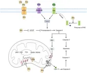
Mitochondria are central to the regulation of apoptosis, the programmed cell death process, which plays a pivotal role in cholestatic liver injury. The review discusses two primary apoptotic pathways influenced by BAs: death receptor-independent and death receptor-dependent pathways.
In death receptor-independent pathways, BAs can directly impair the mitochondrial electron transport chain (ETC), leading to the production of reactive oxygen species (ROS) and oxidative stress. This stress causes the mitochondrial permeability transition pore (mPTP) to open, disrupting the mitochondrial membrane potential and ultimately leading to cell death. The release of cytochrome C from mitochondria into the cytosol triggers the intrinsic pathway of apoptosis, culminating in the activation of caspase enzymes that execute cell death.
In death receptor-dependent pathways, BAs can interact with death receptors on the cell membrane, such as the FAS receptor, to initiate apoptosis. This interaction leads to the formation of a death-inducing signaling complex that activates downstream caspases, further promoting cell death. The review highlights the role of Bcl-2 family proteins, such as BAX and BAK, which facilitate mitochondrial membrane permeabilization during apoptosis.
Endoplasmic Reticulum Stress and Inflammatory Responses
The endoplasmic reticulum (ER) is crucial for protein synthesis, folding, and calcium storage. Cholestasis can trigger ER stress, particularly when misfolded proteins accumulate due to disrupted cellular processes. The review explains how prolonged ER stress leads to the hyperactivation of the unfolded protein response (UPR), which can initiate apoptosis through various pathways, including the activation of pro-apoptotic Bcl-2 proteins and the release of calcium from the ER.
The inflammatory response is another critical aspect of cholestatic liver injury. Although apoptosis is typically an immune-silent process, cholestasis-induced liver injury is often accompanied by significant inflammation. The review details how BAs induce the release of mitochondrial DNA (mtDNA) into the cytosol, where it acts as a damage-associated molecular pattern (DAMP). This mtDNA can activate Toll-like receptor 9 (TLR9), triggering the nuclear factor kappa-B (NF-κB) signaling pathway and leading to the production of inflammatory cytokines. These cytokines promote the recruitment of immune cells, such as neutrophils, to the liver, exacerbating inflammation and liver damage.
Inflammatory Response and Autophagy Dysfunction
Beyond apoptosis, BAs also trigger significant inflammatory responses by releasing mtDNA, which activates TLR9 and leads to the production of pro-inflammatory cytokines. These cytokines contribute to the chemotaxis of neutrophils, key players in the inflammatory response to liver injury. The review discusses how excessive neutrophil infiltration can lead to persistent inflammation, impeding the healing process and exacerbating liver damage.
Autophagy, the process by which cells degrade and recycle damaged organelles and proteins, is another critical pathway affected by cholestasis. The review highlights that cholestasis impairs autophagy by disrupting the fusion of autophagosomes with lysosomes, leading to the accumulation of damaged mitochondria and other cellular debris. This autophagy dysfunction further contributes to liver injury by promoting oxidative stress and inflammation. The review also discusses the role of key regulatory proteins, such as transcription factor EB (TFEB) and Rab7, in maintaining autophagic flux and how their disruption during cholestasis leads to the pathological accumulation of p62 and other autophagic substrates.
Advances in Clinical Research
Recent advances in the clinical management of cholestasis have focused on targeting mitochondrial dysfunction and bile acid metabolism. The review discusses several novel therapeutic agents that have shown promise in treating cholestasis by modulating these pathways. For instance, obeticholic acid (OCA), a potent farnesoid X receptor (FXR) agonist, has demonstrated efficacy in improving liver function in patients with primary biliary cholangitis (PBC) who are inadequate responders to ursodeoxycholic acid (UDCA). However, its use is limited by adverse effects such as pruritus and changes in lipid profiles.
The review also explores emerging therapies that target inflammatory pathways, such as cytokine neutralizers and signal transducers. For example, rituximab, a monoclonal antibody targeting the CD20 antigen on B cells, has shown some potential in improving liver biochemistry in PBC patients. Additionally, the review highlights the importance of mitochondria-targeted therapies, such as cyclosporine A (CsA), which inhibits mPTP opening and protects against mitochondrial dysfunction.
Discussion and Future Prospects
The review concludes by emphasizing the need for further research to fully elucidate the molecular mechanisms linking mitochondrial dysfunction to cholestatic liver injury. While there is substantial evidence supporting the role of mitochondrial pathways in this condition, many mechanistic details remain unclear. Future research should focus on understanding the crosstalk between apoptosis, autophagy, and inflammatory responses in cholestasis. Additionally, the development of more refined experimental tools is necessary to investigate the molecular architecture of the mPTP and its role in liver injury.
The review also calls for the advancement of clinical trials for mitochondria-targeting drugs, which are currently limited. Although preclinical studies have shown promising results, translating these findings into effective therapies for cholestasis remains a significant challenge. Addressing these gaps will be crucial for developing novel therapeutic strategies that can effectively manage cholestatic liver injury and improve patient outcomes.
Conclusions
Cholestasis presents a complex challenge in liver disease, with mitochondrial dysfunction playing a central role in its pathogenesis. This review underscores the importance of targeting mitochondrial pathways in developing new treatments for cholestasis. By advancing our understanding of the molecular mechanisms underlying this condition, we can pave the way for innovative therapies that address the root causes of liver injury, offering hope for improved management of cholestatic liver diseases.
Full text
https://www.xiahepublishing.com/2310-8819/JCTH-2024-00087
The study was recently published in the Journal of Clinical and Translational Hepatology.
The Journal of Clinical and Translational Hepatology (JCTH) is owned by the Second Affiliated Hospital of Chongqing Medical University and published by XIA & HE Publishing Inc. JCTH publishes high quality, peer reviewed studies in the translational and clinical human health sciences of liver diseases. JCTH has established high standards for publication of original research, which are characterized by a study’s novelty, quality, and ethical conduct in the scientific process as well as in the communication of the research findings. Each issue includes articles by leading authorities on topics in hepatology that are germane to the most current challenges in the field. Special features include reports on the latest advances in drug development and technology that are relevant to liver diseases. Regular features of JCTH also include editorials, correspondences and invited commentaries on rapidly progressing areas in hepatology. All articles published by JCTH, both solicited and unsolicited, must pass our rigorous peer review process.
Follow us on X: @xiahepublishing
Follow us on LinkedIn: Xia & He Publishing Inc.
END


