By Glenn Roberts Jr.
An X-ray instrument at Berkeley Lab contributed to a battery study that used an innovative approach to machine learning to speed up the learning curve about a process that shortens the life of fast-charging lithium batteries.
Researchers used Berkeley Lab's Advanced Light Source, a synchrotron that produces light ranging from the infrared to X-rays for dozens of simultaneous experiments, to perform a chemical imaging technique known as scanning transmission X-ray microscopy, or STXM, at a state-of-the-art ALS beamline dubbed COSMIC.
Researchers also employed "in situ" X-ray diffraction at another synchrotron - SLAC's Stanford Synchrotron Radiation Lightsource - which attempted to recreate the conditions present in a battery, and additionally provided a many-particle battery model. All three forms of data were combined in a format to help the machine-learning algorithms learn the physics at work in the battery.
While typical machine-learning algorithms seek out images that either do or don't match a training set of images, in this study the researchers applied a deeper set of data from experiments and other sources to enable more refined results. It represents the first time this brand of "scientific machine learning" was applied to battery cycling, researchers noted. The study was published recently in Nature Materials.
The study benefited from an ability at the COSMIC beamline to single out the chemical states of about 100 individual particles, which was enabled by COSMIC's high-speed, high-resolution imaging capabilities. Young-Sang Yu, a research scientist at the ALS who participated in the study, noted that each selected particle was imaged at about 50 different energy steps during the cycling process, for a total of 5,000 images.
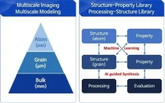
The data from ALS experiments and other experiments were combined with data from fast-charging mathematical models, and with information about the chemistry and physics of fast charging, and then incorporated into the machine-learning algorithms.
"Rather than having the computer directly figure out the model by simply feeding it data, as we did in the two previous studies, we taught the computer how to choose or learn the right equations, and thus the right physics," said Stanford postdoctoral researcher Stephen Dongmin Kang, a study co-author.
Patrick Herring, senior research scientist for Toyota Research Institute, which supported the work through its Accelerated Materials Design and Discovery program, said, "By understanding the fundamental reactions that occur within the battery, we can extend its life, enable faster charging, and ultimately design better battery materials."
Read SLAC's release, "In a leap for battery research, machine learning gets scientific smarts."
To Speed Discovery, Infrared Microscopy Goes 'Off the Grid'
By Lori Tamura
Question: What do a roundworm, a Sharpie pen, and high-vacuum grease have in common? Answer: They've all been analyzed in recent proof-of-principle microscopy experiments at Berkeley Lab's Advanced Light Source (ALS).
In the journal Communications Biology , researchers from Caltech, UC Berkeley, and the Berkeley Synchrotron Infrared Structural Biology Imaging Program (BSISB) reported a more efficient way to collect "high-dimensional" infrared images - where each pixel contains rich physical and chemical information. With the new method, scans that would've taken up to 10 hours to complete can now be done in under an hour, potentially broadening the scope of biological spectromicroscopy to time-sensitive experiments.
"We realized that sampling our model organism - the small roundworm C. elegans - as it changes over time was challenging for software rather than hardware reasons," said Elizabeth Holman, a graduate student in chemistry at Caltech and co-first author of the paper. "For example, image sampling was limited to uniform-grid raster scans with rectangular boundaries and fixed distances between sample points."
The new technique, implemented at the ALS with co-first author Yuan-Sheng Fang, a graduate student in physics at UC Berkeley, uses a grid-less, adaptive approach that autonomously increases sampling in areas displaying greater physical or chemical contrast. In the proof-of-concept infrared microscopy experiments, the researchers examined two samples.
The first was a two-component system in which both components (permanent-marker ink and high-vacuum grease) were well characterized. Details of the sample were very difficult to see clearly with the naked eye, so it was a good test of how the software would perform with minimal guidance from a human experimenter. The second sample was a live, larval-stage C. elegans, a biological model system studied by thousands of researchers.
In both cases, autonomous adaptive data acquisition (AADA) methods clearly outperformed nonadaptive methods. In the second example, increased sampling density corresponded with known C. elegans anatomical features, and the head region was mapped in 45 minutes versus about 4.9 hours using commercially available software.
"Outside of our specific published work, the results suggest that integrating AADA into existing scanning-based satellite, drone, and/or microscope techniques can facilitate research in fields ranging from hyperspectral remote sensing to ocean and space exploration," said Holman.
Actor in a Supporting Role: Substrate Effects on 2D Layers
By Lori Tamura
Atomically thin layers are of great technological interest because of potentially useful electronic properties that emerge as the layer thickness approaches the 2D limit. Such materials tend to form weak bonds outside the layer and are thus generally assumed to be unaffected by substrates that provide physical support.
To make further progress, however, scientists must rigorously test this assumption, not only to better understand single-layer physics, but also because the existence of substrate effects raises the possibility of tuning layer properties by tweaking the substrate.
As reported in the journal Physical Review Letters, a team led by Tai-Chang Chiang of the University of Illinois at Urbana-Champaign and his postdoctoral associate, Meng-Kai Lin, used Berkeley Lab's Advanced Light Source (ALS) to probe changes in the electronic properties of a 2D semiconductor, titanium telluride, as the thickness of a substrate, platinum telluride, was increased. Single-layer titanium telluride is highly sensitive to what lies underneath, making it particularly useful as a test case for investigating substrate coupling effects.
The results showed that as the substrate thickness increased, a dramatic and systematic variation occurred in the single-layer titanium telluride. An electronic phenomenon known as a charge density wave -- a coupled charge and lattice distortion characteristic of single-layer titanium telluride -- was suppressed.
"The experimental findings, combined with first-principles theoretical simulations, led to a detailed explanation of the results in terms of the basic quantum mechanical interactions between the single layer and the tunable substrate," said Lin.
Given that the interfacial bonding remained weak, the researchers concluded that the observed changes were correlated with the substrate's transformation from a semiconductor to a semimetal as it increased in thickness.
"This systematic study illustrates the crucial role that substrate interactions play in the physics of ultrathin films," said Lin. "The scientific understanding derived from our work also provides a framework for designing and engineering ultrathin films for useful and enhanced properties."
INFORMATION:
