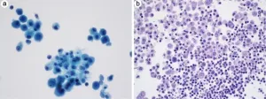
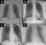
Serous effusion, defined as the excessive accumulation of fluid in body cavities such as the pleural, pericardial, and peritoneal spaces, is a critical diagnostic challenge in pathology. Cytological evaluation of serous fluids provides vital information for detecting underlying etiologies, such as malignancy, and helps in evaluating tumor stages and customizing treatment plans. To address inconsistencies in the diagnostic criteria and nomenclature used in fluid cytology reporting, the International Academy of Cytology and the American Society of Cytopathology introduced The International System (TIS) for reporting serous fluid cytopathology. This system, published in December 2020, aims to standardize diagnostic criteria, improve reproducibility of reports, and enhance communication between pathologists and clinicians. The TIS framework categorizes serous fluid samples into five distinct diagnostic categories: Nondiagnostic (ND), Negative for Malignancy (NFM), Atypia of Uncertain Significance (AUS), Suspicious for Malignancy (SFM), and Malignant (MAL). This review provides a comprehensive summary of the TIS categories, their respective risks of malignancy (ROM), potential diagnostic pitfalls, and a practical approach to serous fluid specimen analysis.
Nondiagnostic (ND)
The ND category includes specimens that fail to provide sufficient diagnostic information, typically due to inadequate cellularity, extensive degeneration, or excessive blood contamination. Samples are classified as ND when they contain fewer than ten cells, such as benign lymphocytes or macrophages, or when the sample is primarily composed of red blood cells. Adequate sample volume is crucial for a reliable diagnosis, with at least 75–100 mL recommended for optimal detection of malignancies. However, smaller volumes should not be outright rejected if they contain adequate cellular material. Even in cases with a suitable volume, samples can be deemed ND if the cells exhibit extensive degenerative changes or if the sample lacks diagnostic cells such as mesothelial cells.
Negative for Malignancy (NFM)
Specimens classified as NFM demonstrate cellular changes that are entirely devoid of malignant characteristics. This category includes samples with reactive or benign changes, such as mesothelial hyperplasia, chronic inflammation, or benign lymphocytic predominance. The NFM category is crucial in clinical scenarios where malignancy is suspected, as it can guide further diagnostic or therapeutic actions. The ROM for NFM varies widely, and while most cases are truly negative, rare instances of false negatives can occur, underscoring the need for thorough clinical correlation.
Atypia of Uncertain Significance (AUS)
The AUS category is reserved for specimens that exhibit limited cellular or architectural atypia that does not clearly indicate malignancy. This category includes cases with mild nuclear abnormalities, such as slight nuclear enlargement or hyperchromasia, or architectural features like papillary clusters that are suspicious but not definitive for malignancy. AUS is a challenging category as it often requires careful consideration of the clinical context and may necessitate additional sampling or ancillary studies, such as immunohistochemistry, to clarify the diagnosis. The ROM for AUS is variable, reflecting the heterogeneous nature of cases included in this category.
Suspicious for Malignancy (SFM)
The SFM category includes specimens that show cytological features suspicious for malignancy but fall short of a definitive malignant diagnosis. These cases often display significant cytological atypia, such as irregular nuclear contours, coarse chromatin, or prominent nucleoli, but lack confirmatory evidence of malignancy. The SFM classification necessitates a cautious approach, as these cases have a high ROM, often approaching that of definitive malignancies. Management of SFM cases typically involves more aggressive follow-up, including repeat sampling or additional diagnostic procedures.
Malignant (MAL)
The MAL category is assigned to specimens with definitive cytomorphological or ancillary study findings diagnostic of malignancy. This includes both primary malignancies, such as malignant mesothelioma, and secondary malignancies like metastatic carcinoma, which is the most common cause of malignant effusions. Malignant effusions often arise from primary cancers of the lung, breast, ovary, or gastrointestinal tract, among others. The ROM for the MAL category is nearly 100%, highlighting the importance of accurate cytological assessment in guiding patient management.
Conclusions
The TIS for reporting serous fluid cytopathology represents a significant advancement in the standardization of diagnostic criteria and nomenclature in fluid cytology. By providing a clear and consistent framework, TIS enhances the reproducibility of cytopathology reports, improves communication between pathologists and clinicians, and offers valuable guidance for patient management. The five-tiered system—ND, NFM, AUS, SFM, and MAL—each with specific diagnostic criteria and associated ROM, serves as a comprehensive tool for the accurate assessment of serous fluid specimens. Future studies and clinical experience will continue to refine the application and interpretation of TIS, ensuring its relevance and effectiveness in diverse clinical settings.
Full text
https://www.xiahepublishing.com/2771-165X/JCTP-2023-00025
The study was recently published in the Journal of Clinical and Translational Pathology.
Journal of Clinical and Translational Pathology (JCTP) is the official scientific journal of the Chinese American Pathologists Association (CAPA). It publishes high quality peer-reviewed original research, reviews, perspectives, commentaries, and letters that are pertinent to clinical and translational pathology, including but not limited to anatomic pathology and clinical pathology. Basic scientific research on pathogenesis of diseases as well as application of pathology-related diagnostic techniques or methodologies also fit the scope of the JCTP.
Follow us on X: @xiahepublishing
Follow us on LinkedIn: Xia & He Publishing Inc.
END


