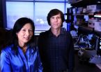
(Press-News.org) A corrective strategy used by astronomers to sharpen images of celestial bodies can now help scientists see with more depth and clarity into the living brain of a mouse. Eric Betzig, a group leader at the Howard Hughes Medical Institute's Janelia Farm Research Campus, will present his team's latest work using adaptive optics for biology at the annual meeting of the American Association for the Advancement of Science in Washington, D.C. during a press conference on Thursday, Feb., 17, and a panel discussion on Friday, Feb. 18.
A key problem in microscopy is that when the light shines on a biological sample, such as a slice of brain tissue, light waves hit the cells and bounce off in different directions. The larger the piece of tissue, the more interesting and diverse its collection of parts, which makes the light waves bend and scatter in unpredictable ways.
For the past decade, researchers have been trying, with limited success, to sharpen blurred images of biological specimens using a method astronomers call adaptive optics. Recently, however, Betzig and postdoctoral researcher Na Ji, have made large strides toward improving resolution deep into tissue by combining a new approach to adaptive optics with an imaging technology called two photon fluorescence microscopy. Their results, published in 2009 in the journal Nature Methods, describe the first applications of adaptive optics to improve images of brain slices taken from mice.
Astronomers apply adaptive optics by shining a laser high in the atmosphere in the same direction as the star or other object they want to observe. The light returning from this so-called guide star gets distorted as it travels through the turbulent atmosphere back to the telescope. By using a tool called a wavefront sensor, astronomers can measure this distortion directly, and then use these measurements to deform a telescope mirror to cancel out the atmospheric aberrations. The correction gives a much clearer view of the target. Today, adaptive optics instrumentation accompanies all of the world's major telescopes.
Unlike in astronomy, microscopists can't place a wavefront sensor within a live animal to directly measure the distortions of light deep within tissue. To get around this problem, Betzig reasoned that the perfect focus is nothing more than a bunch of rays converging from many different directions to the same identical point. The heterogeneity of tissue means that ray is deflected differently so they no longer meet at a single point. Betzig figured that if they could study the rays individually, they could correct their deflections and steer them back to a single focus.
For their 2009 study, Betzig and Ji buried fluorescent beads underneath thick slices of mouse brain. The beads act as guide stars to help measure the deflections of the rays. To do that, the pair use a one-inch display called a spatial light modulator. The display allows them to turn on one ray at a time and then take an image of the bead. They can then determine how much the ray is deflected from the amount the bead's image is shifted relative to the desired focal point. The display is then used like a small, tiltable mirror to steer the ray back to the focal point. The process is then repeated with each of the other rays. This strategy improves the fluorescent signal and recovers optimal resolution through a chunk of tissue up to 400 micrometers thick. "Another advantage is that it's very efficient in terms of how little light is needed," Betzig says. "Light is not completely noninvasive, so as microscopists we have to be very careful not to damage our specimens."
Ji, who will soon begin her own lab as a fellow at Janelia Farm, has recently taken the technique to the next level: live brain imaging in mice. To do this, she uses genetic engineering to label the brains of mouse fetuses with a fluorescent marker of neurons while at the same time injecting fluorescent bead guide stars. After the mice mature, Ji builds a window into the brain by removing a piece of skull and replacing it with a clear glass cover. She then uses the adaptive optical microscope she and Betzig built to peer through the glass and image the individual neurons underneath.
One of the concerns the researchers faced when imaging a live animal was how long a correction would persist. In astronomy, stars twinkle so fast that up to a thousand corrections are needed per second. Fortunately, Ji and Betzig learned that, for an anesthetized mouse, a single correction would remain valid for nearly an hour. Another question was whether the correction made at a single guide star bead could be applied to surrounding areas. Although the answer varies from sample to sample, Ji's work has shown that a single correction will often apply to a space of over 100 micrometers in each direction, a volume that can fit dozens of neurons. "It would be prohibitively slow if you had to correct at every point throughout an entire volume," Betzig says. To address this same problem in the field of astronomy, scientists employ a series of deformable mirrors that each look at guide stars in different directions. Betzig hopes to use a similar strategy to widen the correction even further in his microscope.
Since arriving at Janelia Farm in 2005, Betzig and his colleagues have pioneered new super high-resolution imaging techniques and shared them with biologists. One of them, photoactivated localization microscopy or PALM, maps individual protein molecules to produce images with 10-20 times the resolution of a traditional light microscope. PALM and other types of imaging, such as confocal microscopy and wide-field imaging, can be greatly improved with adaptive optics, he says. This is only the beginning. "What we do is primitive compared to the sophistication of what they do in the astronomy community," Betzig says. "I still feel we have a lot to learn from astronomers."
###
Press conference: 2 p.m. EST on Thursday, February 17, 2011, AAAS Newsroom Headquarters, Room 204A on the second level of the Washington Convention Center.
Panel Discussion: Sharper Images in Astronomy, Microscopy, and Vision Science Using Adaptive Optics, Friday, February 18, 2011: 10:00 a.m.-11:30 a.m., room 101, Washington Convention Center
Contacts
Andrea Widener, 301-215-8807, widenera@hhmi.org
Jim Keeley, 301-215-8858, keeleyj@hhmi.org
The Howard Hughes Medical Institute plays a powerful role in advancing scientific research and education in the United States. Its scientists, located across the country and around the world, have made important discoveries that advance both human health and our fundamental understanding of biology. The Institute also aims to transform science education into a creative, interdisciplinary endeavor that reflects the excitement of real research. For more information, visit www.hhmi.org.
END
Plants have for the first time been cloned as seeds. The research by aUC Davis plant scientists and their international collaborators, published Feb. 18 in the journal Science, is a major step towards making hybrid crop plants that can retain favorable traits from generation to generation.
Most successful crop varieties are hybrids, said Simon Chan, assistant professor of plant biology at UC Davis and an author of the paper. But when hybrids go through sexual reproduction, their traits, such as fruit size or frost resistance, get scrambled and may be lost.
"We're ...
Scientists have identified the gene that allows the Rubella virus to block cell death and reverse engineered a mutant gene that slows the virus's spread. Tom Hobman and a team of researchers at the University of Alberta's Faculty of Medicine and Dentistry believed that RNA viruses were able to spread by blocking the pathways in cells that lead to cell suicide, and isolated the responsible gene in Rubella, also known as German measles. They then created a mutant version of this gene that made the virus spread more slowly. These results are reported in PLoS Pathogens.
The ...
MADISON — Most people cross borders such as doorways or state lines without thinking much about it. Yet not all borders are places of limbo intended only for crossing. Some borders, like those between two materials that are brought together, are dynamic places where special things can happen.
For an electron moving from one material toward the other, this space is where it can join other electrons, which together can create current, magnetism or even light.
A multi-institutional team has made fundamental discoveries at the border regions, called interfaces, between ...
Glaucoma – a leading cause of vision loss and blindness worldwide – runs in families. A team of investigators from Vanderbilt University and the University of Florida has identified a new candidate gene for the most common form of the eye disorder, primary open angle glaucoma (POAG).
The findings, reported Feb. 17 in the open-access journal PLoS Genetics, offer novel insights into glaucoma pathology and could lead to targeted treatment strategies.
Elevated pressure inside the eye is a strong risk factor for POAG. Pressure increases because of increased resistance to the ...
Cases of ventilator-associated pneumonia — the most lethal and among the most common of all hospital-associated infections — dropped by more than 70 percent in Michigan hospitals where medical staff used a simple checklist designed by Johns Hopkins researchers. Such pneumonias kill an estimated 36,000 Americans each year.
The findings, published online in the journal Infection Control and Hospital Epidemiology, show how a relatively simple series of steps, coupled with an education program and a culture that promotes patient safety, can save tens of thousands of lives ...
PITTSBURGH, Feb. 17 – Researchers at the University of Pittsburgh have been awarded funding for two projects that will place brain-computer interfaces (BCI) in patients with spinal cord injuries to test if it is possible for them to control external devices, such as a computer cursor or a prosthetic limb, with their thoughts.
The projects build upon ongoing research conducted in epilepsy patients who had the interfaces temporarily placed on their brains and were able to move cursors and play computer games, as well as in monkeys that through interfaces guided a robotic ...
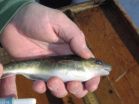
NEW YORK, Feb. 17, 2011 – A research group led by a New York University School of Medicine scientist discovered a genetic variant that allows a fish in the Hudson River to live in waters heavily polluted by PCBs. In a study published in the February 18, 2011, online issue of Science, they report that a population of Hudson River fish apparently evolved rapidly in response to the toxic chemicals, which were first introduced in 1929, and were banned fifty years later. PCBs, or polychlorinated biphenyls, were used in hundreds of industrial and commercial applications, especially ...
(Millbrook, N.Y. – Feb 17, 2011) Forest biomass could replace as much as one quarter of the liquid fossil fuel now being used for industrial and commercial heating in the Northeastern United States. That's according to a new report released today by the Cary Institute of Ecosystem Studies.
But the report also has sharp caveats: The potential for forest biomass varies widely within the region, and forest resources must be carefully managed to protect the other important services and goods they provide. Under the right circumstances, however, the report found that forest ...
Researchers at the University of California, San Diego School of Medicine have identified the motors that move non-infectious prion proteins (PrPC) – found within many mammalian cells – up and down long, neuronal transport pathways. Identifying normal movement mechanisms of PrPC may help researchers understand the spread of infectious prions within and between neurons to reach the brain, and aid in development of therapies to halt the transport.
Their study is published in the February 18 edition of the journal Cell.
The small prion protein is found in the cell membrane ...
For 30 years, two General Electric facilities released about 1.3 million pounds of polychlorinated biphenyls (PCBs) into New York's Hudson River, devastating and contaminating fish populations. Some 50 years later, one type of fish—the Atlantic tomcod—has not only survived but appears to be thriving in the hostile Hudson environment.
Researchers from Woods Hole Oceanographic Institution (WHOI) have joined colleagues from New York University (NYU) and NOAA to investigate this phenomenon and report that the tomcod living in the Hudson River have undergone a rapid evolutionary ...


