(Press-News.org) Pancreatic cancer is a particularly devastating disease. At least 94 percent of patients will die within five years, and in 2013 it was ranked as one of the top 10 deadliest cancers.
Routine screenings for breast, colon and lung cancers have improved treatment and outcomes for patients with these diseases, largely because the cancer can be detected early. But because little is known about how pancreatic cancer behaves, patients often receive a diagnosis when it's already too late.
University of Washington scientists and engineers are developing a low-cost device that could help pathologists diagnose pancreatic cancer earlier and faster. The prototype can perform the basic steps for processing a biopsy, relying on fluid transport instead of human hands to process the tissue. The team presented its initial results this month (February 2014) at the SPIE Photonics West conference and recently filed a patent for this first-generation device and future technology advancements.
"This new process is expected to help the pathologist make a more rapid diagnosis and be able to determine more accurately how invasive the cancer has become, leading to improved prognosis," said Eric Seibel, a UW research professor of mechanical engineering and director of the department's Human Photonics Laboratory.
The new instrumentation would essentially automate and streamline the manual, time-consuming process a pathology lab goes through to diagnose cancer. Currently, a pathologist takes a biopsy tissue sample, then sends it to the lab where it's cut into thin slices, stained and put on slides, then analyzed optically in 2-D for abnormalities.
The UW's technology would process and analyze whole tissue biopsies for 3-D imaging, which offers a more complete picture of the cellular makeup of a tumor, said Ronnie Das, a UW postdoctoral researcher in bioengineering who is the lead author on a related paper.
"As soon as you cut a piece of tissue, you lose information about it. If you can keep the original tissue biopsy intact, you can see the whole story of abnormal cell growth. You can also see connections, cell morphology and structure as it looks in the body," Das said.
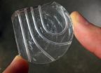
The research team is building a thick, credit card-sized, flexible device out of silicon that allows a piece of tissue to pass through tiny channels and undergo a series of steps that replicate what happens on a much larger scale in a pathology lab. The device harnesses the properties of microfluidics, which allows tissue to move and stop with ease through small channels without needing to apply a lot of external force. It also keeps clinicians from having to handle the tissue; instead, a tissue biopsy taken with a syringe needle could be deposited directly into the device to begin processing.
Researchers say this is the first time material larger than a single-celled organism has successfully moved in a microfluidic device. This could have implications across the sciences in automating analyses that usually are done by humans.
Das and Chris Burfeind, a UW undergraduate student in mechanical engineering, designed the device to be simple to manufacture and use. They first built a mold using a petri dish and Teflon tubes, then poured a viscous, silicon material into the mold. The result is a small, transparent instrument with seamless channels that are both curved and straight.
The researchers have used the instrument to process a tissue biopsy one step at a time, following the same steps as a pathology lab would. Next, they hope to combine all of the steps into a more robust device – including 3-D imaging – then build and optimize it for use in a lab. Future iterations of the device could include layers of channels that would allow more analyses on a piece of tissue without adding more bulk to the device.
The UW researchers say the technology could be used overseas as an over-the-counter kit that would process biopsies, then send that information to pathologists who could look for signs of cancer from remote locations. Additionally, it could potentially reduce the time it takes to diagnose cancer to a matter of minutes, Das said.
INFORMATION:
The team is working with Melissa Upton, a pathologist with UW Medicine. The research is funded by the National Science Foundation Bioengineering division and the U.S. Department of Education Graduate Assistance in Areas of National Need program.
For more information, contact Seibel at eseibel@uw.edu or 206-616-1486, and Das at rdas@uw.edu or 206-221-3813.
Grant numbers: NSF Bioengineering division (CBET-1212540).
Posted with photos, videos: http://www.washington.edu/news/2014/02/06/credit-card-sized-device-could-analyze-biopsy-help-diagnose-pancreatic-cancer-in-minutes/
Credit card-sized device could analyze biopsy, help diagnose pancreatic cancer in minutes
2014-02-06
ELSE PRESS RELEASES FROM THIS DATE:
UI researchers evaluate best weather forecasting models
2014-02-06
Two University of Iowa researchers recently tested the ability of the world's most advanced weather forecasting models to predict the Sept. 9-16, 2013 extreme rainfall that caused severe flooding in Boulder, Colo.
The results, published in the December 2013 issue of the journal Geophysical Research Letters, indicated the forecasting models generally performed well, but also left room for improvement.
David Lavers and Gabriele Villarini, researchers at IIHR—Hydroscience and Engineering, a world-renowned UI research facility, evaluated rainfall forecasts from eight different ...
Nanoparticle pinpoints blood vessel plaques
2014-02-06
A team of researchers, led by scientists at Case Western Reserve University, has developed a multifunctional nanoparticle that enables magnetic resonance imaging (MRI) to pinpoint blood vessel plaques caused by atherosclerosis. The technology is a step toward creating a non-invasive method of identifying plaques vulnerable to rupture–the cause of heart attack and stroke—in time for treatment.
Currently, doctors can identify only blood vessels that are narrowing due to plaque accumulation. A doctor makes an incision and slips a catheter inside a blood vessel in the arm, ...
Loose coupling between calcium channels and sensors
2014-02-06
This news release is available in German. Information transmission at the synapse between neurons is a highly complex, but at the same time very fast, series of events. When a voltage change, the so-called action potential, reaches the synaptic terminal in the presynaptic neuron, calcium flows through voltage-gated calcium channels into the presynaptic neuron. This influx leads to a rise in the intracellular calcium concentration. Calcium then binds to a calcium sensor in the presynaptic terminal, which in turn triggers the release of vesicles containing neurotransmitters ...
US lead in science and technology shrinking
2014-02-06
The United States' (U.S.) predominance in science and technology (S&T) eroded further during the last decade, as several Asian nations--particularly China and South Korea--rapidly increased their innovation capacities. According to a report released today by the National Science Board (NSB), the policy making body of the National Science Foundation (NSF) and an advisor to the President and Congress, the major Asian economies, taken together, now perform a larger share of global R&D than the U.S., and China performs nearly as much of the world's high-tech manufacturing as ...
Prickly protein
2014-02-06
A genetic mechanism that controls the production of a large spike-like protein on the surface of Staphylococcus aureus (staph) bacteria alters the ability of the bacteria to form clumps and to cause disease, according to a new University of Iowa study.
The new study is the first to link this genetic mechanism to the production of the giant surface protein and to clumping behavior in bacteria. It is also the first time that clumping behavior has been associated with endocarditis, a serious infection of heart valves that kills 20,000 Americans each year. The findings were ...
NASA study points to infrared-herring in apparent Amazon green-up
2014-02-06
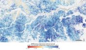
For the past eight years, scientists have been working to make sense of why some satellite data seemed to show the Amazon rain forest "greening-up" during the region's dry season each year from June to October. The green-up indicated productive, thriving vegetation in spite of limited rainfall.
Now, a new NASA study published today in the journal Nature shows that the appearance of canopy greening is not caused by a biophysical change in Amazon forests, but instead by a combination of shadowing within the canopy and the way that satellite sensors observe the Amazon during ...
Valentine's Day advice: Don't let rocky past relations with parents spoil your romance
2014-02-06
University of Alberta relationship researcher Matt Johnson has some Valentine's Day advice for anybody who's had rocky relations with their parents while growing up: don't ...
Falcon feathers pop-up during dive
2014-02-06
Similar to wings and fins with self-adaptive flaps, the feathers on a diving peregrine falcon's feathers may pop-up during high speed dives, according to a study published in PLOS ONE on February 5, 2014 by Benjamin Ponitz from the Institute of Mechanics ...
New, high-tech prosthetics and orthotics offer active life-style for users
2014-02-06
TAMPA, Fla. (Feb. 5, 2014) – Thanks to advanced technologies, those who wear prosthetic and orthotic devices ...
University of Montana research shows converting land to agriculture reduces carbon uptake
2014-02-06
MISSOULA – University of Montana researchers examined the impact that converting natural land to cropland has on global vegetation growth, as measured by satellite-derived ...


