(Press-News.org) VIDEO:
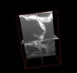
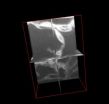
This shows 3-D ortho-rotation of leaf mesophyll cells. Micrographs were collected by milling fixed tissue accompanied by SEM imaging using FIB-SEM. The complete videos published with the article are available...
Click here for more information.
Plant cells are beginning to look a lot different to Dr. A. Bruce Cahoon and his colleagues at Middle Tennessee State University (MTSU). They've adopted a new approach that combines the precision of an ion beam with the imaging capabilities of an electron beam to zoom in at micron-level resolution. Scientists who work with nanodevices have used focused ion beam–scanning electron microscopy (FIB-SEM) for decades, but it is only recently that biologists have begun to explore its capabilities. The researchers at MTSU are the first to optimize its use for plant cell imaging.
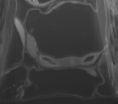
Their results are beautiful grayscale mosaics of plant organelles. Large, dark vacuoles bordered by light, oval-shaped chloroplasts resting along thick, smooth cell walls. Plump berry-like lipid bodies surrounding the round nucleus. Endoplasmic reticulum, protein bodies, and other uniquely shaped structures squeezing together like flexible tiles within the cells. The researchers produced a stunning selection of images as they developed methods to work with seed, leaf, stem, root, and petal cell types of Arabidopsis thaliana. Cahoon and his colleagues describe their methods and results, along with 3D renderings and video, in the June issue of Applications in Plant Sciences.
"We attended an open presentation about FIB-SEM held at MTSU about its capabilities, and we immediately thought of using it on plant tissues," explains Cahoon. The ion beam slices thin sections of a sample, which are each captured in an image by the scanning electron beam. The images of over 100 sections, just nanometers thick, are combined to construct a richly detailed three-dimensional figure.
"Certain aspects were very attractive, but it did mean we would have to step back and innovate the technology for plant tissues," says Cahoon. Fortunately, the researchers were able to benefit from the work of a few other biologists who had begun using FIB-SEM with animal tissues. The animal tissue protocol served as a base from which to modify specific plant techniques.
"The 3D visualization of subcellular structures only seen in 2D views was very satisfying," says Cahoon. "Some were just as you would imagine but others were surprisingly organized—for example, amorphous aggregates in the petal cell vacuoles that had been dismissed in previous electron microscopy studies as artifacts or uninteresting. The 3D view of these structures revealed a regular pattern appearing in almost every petal mesophyll cell used in our study. When molecules are organized like this, it suggests function and poses new questions."
Electron microscopes can produce images at a much higher magnification and resolution than optical light microscopes because electrons have shorter wavelengths than visible light. The ion beam is able to penetrate the tissue sample and slice off thin sections at a precision much greater than any form of mechanical milling. Combining these two technologies offers unique images unattainable by any other method.
Unfortunately, all good things tend to have their drawbacks and FIB-SEM is no exception. It is a time-intensive process that requires an expensive specialized instrument. Tissue samples must undergo highly specific fixation procedures to stabilize them for imaging in the electron microscope, and non-conductive biological cells require a conductive coating, such as platinum or a gold alloy.
For the research team at MTSU, the time-consuming procedures and expensive equipment are well worth the results. "We have new specific questions to address that arose from the expanded view of the internal structures. In addition, now that we know how to use this technology, we have begun to expand the repertoire of photosynthetic organisms to, at least initially, explore their cellular architecture just to see where it takes us."
The new FIB-SEM methods developed for seed, leaf, stem, root, and petal cell types will help expand the toolset available to plant anatomists for understanding the nature of organelles, cells, and plant development. A unique view of plant cell interiors could reveal never-before-seen aspects of the architecture and distribution of organelles. This work is just one example of how technological advances in one field of science, in this case, materials science, can open new doors for researchers in other fields.
INFORMATION:
Bhawana, Joyce L. Miller, and A. Bruce Cahoon. 3D Plant cell architecture of Arabidopsis thaliana (Brassicaceae) using focused ion beam–scanning electron microscopy. Applications in Plant Sciences 2(6): 1300090. doi:10.3732/apps.1300090
Applications in Plant Sciences (APPS) is a monthly, peer-reviewed, open access journal focusing on new tools, technologies, and protocols in all areas of the plant sciences. It is published by the Botanical Society of America, a nonprofit membership society with a mission to promote botany, the field of basic science dealing with the study and inquiry into the form, function, development, diversity, reproduction, evolution, and uses of plants and their interactions within the biosphere. APPS is available as part of BioOne's Open Access collection.
For further information, please contact the APPS staff at apps@botany.org.
Viewing plant cells in 3-D (no glasses required)
Researchers apply FIB-SEM technology to 3-D plant cell architecture imaging
2014-06-09
ELSE PRESS RELEASES FROM THIS DATE:
Needle biopsy underused in breast cancer diagnosis, negatively impacting diagnosis and care
2014-06-09
Needle biopsy, the standard of care radiological procedure for diagnosing breast cancer, is underused with too many patients undergoing the more invasive, excisional biopsy to detect their disease, according to research from The University of Texas MD Anderson Cancer Center.
The study, published in the Journal of Clinical Oncology, also finds that patients are often influenced by surgeons to undergo the unnecessary surgery -- a decision that's costly and can negatively impact their diagnosis and treatment.
A needle biopsy is a non-surgical procedure typically performed ...
Women and health-care providers differ on what matters most about contraception
2014-06-09
LEBANON, NH – When women are choosing a contraceptive, health care providers should be aware that the things they want to discuss may differ from what women want to hear, according to a survey published in the recent issue of the journal Contraception.
Most of the information women receive about contraceptives focuses heavily on the effectiveness in preventing pregnancy, but this information was ranked fifth in importance by women, according to the study conducted by researchers at Dartmouth College.
The researchers conducted an online survey of 417 women, aged 15-45, ...
JCI online ahead of print contents for June 9, 2014
2014-06-09
Clinical trial evaluates ex vivo cultured cord blood
Umbilical cord blood (UCB) is a rich source of hematopoietic stem and progenitor cells (HSPCs) that can be used for bone marrow transplantation; however, UCB transplantation is hampered by low numbers of HSPCs per donation, which delays engraftment and immune reconstitution. In this issue of the Journal of Clinical Investigation, Mitchell Horwitz and colleagues at Duke University Medical Center conducted a phase I clinical trial to test the long term engraftment capability of UCB HSPCs that were expanded ex vivo for ...
Newly identified B-cell selection process adds to understanding of antibody diversity
2014-06-09
BOSTON – As elite soldiers of the body's immune response, B cells serve as a vast standing army ready to recognize and destroy invading antigens, including infections and cancer cells. To do so, each new B cell comes equipped with its own highly specialized weapon, a unique antibody protein that selectively binds to specific parts of the antigen. The key to this specialization is the antigen-binding region that tailors each B cell to a particular antigen, determining whether B cells survive boot camp and are selected for maturation and survival, or wash out and die.
Now, ...
Faster, higher, stronger: A protein that enables powerful initial immune response
2014-06-09
Your first response to an infectious agent or antigen ordinarily takes about a week, and is relatively weak. However, if your immune system encounters that antigen a second time, the so-called memory response is rapid, powerful, and very effective.
Now, a team of researchers at The Wistar Institute offers evidence that a protein, called Foxp1, is a key component of these antibody responses. Manipulating this protein's activity, they say, could provide a useful pathway to boosting antibody responses to treat infectious diseases, for example, or suppressing them to treat ...
The Academy of Radiology Research featured in Nature Biotechnology journal
2014-06-09
The Academy of Radiology Research reported in the current issue of Nature Biotechnology (Volume 32, Issue 6) that patent output from the National Institutes of Health (NIH) is vital to understanding which various areas of science are contributing most to America's innovation economy. The report, "Patents as Proxies: NIH Hubs of Innovation," confirms an increased economic value of NIH patents as compared to private sector patents, as well as meaningful differences in the rate and quality of invention across different research and development (R&D) investments.
"The Academy ...
NOAA scientists find mosquito control pesticide low risk to juvenile oysters, hard clams
2014-06-09
Four of the most common mosquito pesticides used along the east and Gulf coasts show little risk to juvenile hard clams and oysters, according to a NOAA study.
However, the study, published in the on-line journal Archives of Environmental Contamination and Toxicology, also determined that lower oxygen levels in the water, known as hypoxia, and increased acidification actually increased how toxic some of the pesticides were. Such climate variables should be considered when using these pesticides in the coastal zone, the study concluded.
"What we found is that larval oysters ...
Antiviral therapy may prevent liver cancer in hepatitis B patients
2014-06-09
DETROIT – Researchers have found that antiviral therapy may be successful in preventing hepatitis B virus from developing into the most common form of liver cancer, hepatocellular carcinoma (HCC).
That was the finding of a study published in the May issue of Clinical Gastroenterology and Hepatology. Investigators from Henry Ford Health System in Detroit, Geisinger Health System in Danville, Pa., and Kaiser Permanente in Honolulu, Hawaii and Portland, Ore. participated in the study, along with investigators from the Centers for Disease Control and Prevention in Atlanta. ...
How much fertilizer is too much for the climate?
2014-06-09
EAST LANSING, Mich. — Helping farmers around the globe apply more-precise amounts of nitrogen-based fertilizer can help combat climate change.
In a new study published in this week's Proceedings of the National Academy of Sciences, Michigan State University researchers provide an improved prediction of nitrogen fertilizer's contribution to greenhouse gas emissions from agricultural fields.
The study uses data from around the world to show that emissions of nitrous oxide, a greenhouse gas produced in the soil following nitrogen addition, rise faster than previously expected ...
Coral, human cells linked in death
2014-06-09
SAN DIEGO (June 6, 2014) — Humans and corals are about as different from one another as living creatures get, but a new finding reveals that in one important way, they are more similar than anyone ever realized.
A biologist at San Diego State University has discovered they share the same biomechanical pathway responsible for triggering cellular self-destruction. That might sound scary, but killing off defective cells is essential to keeping an organism healthy.
The finding will help biologists advance their understanding of the early evolution of multicellular life, ...
LAST 30 PRESS RELEASES:
New knowledge on heritability paves the way for better treatment of people with chronic inflammatory bowel disease
Under the Lens: Microbiologists Nicola Holden and Gil Domingue weigh in on the raw milk debate
Science reveals why you can’t resist a snack – even when you’re full
Kidney cancer study finds belzutifan plus pembrolizumab post-surgery helps patients at high risk for relapse stay cancer-free longer
Alkali cation effects in electrochemical carbon dioxide reduction
Test platforms for charging wireless cars now fit on a bench
$3 million NIH grant funds national study of Medicare Advantage’s benefit expansion into social supports
Amplified Sciences achieves CAP accreditation for cutting-edge diagnostic lab
Fred Hutch announces 12 recipients of the annual Harold M. Weintraub Graduate Student Award
Native forest litter helps rebuild soil life in post-mining landscapes
Mountain soils in arid regions may emit more greenhouse gas as climate shifts, new study finds
Pairing biochar with other soil amendments could unlock stronger gains in soil health
Why do we get a skip in our step when we’re happy? Thank dopamine
UC Irvine scientists uncover cellular mechanism behind muscle repair
Platform to map living brain noninvasively takes next big step
Stress-testing the Cascadia Subduction Zone reveals variability that could impact how earthquakes spread
We may be underestimating the true carbon cost of northern wildfires
Blood test predicts which bladder cancer patients may safely skip surgery
Kennesaw State's Vijay Anand honored as National Academy of Inventors Senior Member
Recovery from whaling reveals the role of age in Humpback reproduction
Can the canny tick help prevent disease like MS and cancer?
Newcomer children show lower rates of emergency department use for non‑urgent conditions, study finds
Cognitive and neuropsychiatric function in former American football players
From trash to climate tech: rubber gloves find new life as carbon capturers materials
A step towards needed treatments for hantaviruses in new molecular map
Boys are more motivated, while girls are more compassionate?
Study identifies opposing roles for IL6 and IL6R in long-term mortality
AI accurately spots medical disorder from privacy-conscious hand images
Transient Pauli blocking for broadband ultrafast optical switching
Political polarization can spur CO2 emissions, stymie climate action
[Press-News.org] Viewing plant cells in 3-D (no glasses required)Researchers apply FIB-SEM technology to 3-D plant cell architecture imaging


