(Press-News.org) In 2020, the International Agency for Research on Cancer of the World Health Organization stated that breast cancer accounts for most cancer morbidities and mortalities in women worldwide. This alarming statistic not only necessitates newer methods for the early diagnosis of breast cancer, but also brings to light the importance of risk prediction of the occurrence and development of this disease. Ultrasound is an effective and noninvasive diagnostic procedure that truly saves lives; however, it is sometimes difficult for ultrasonologists to distinguish between malignant tumors and other types of benign growths. In particular, in China, breast masses are classified into four categories: benign tumors, malignant tumors, inflammatory masses, and adenosis (enlargement of milk-producing glands). When a benign breast mass is misdiagnosed as a malignant tumor, a biopsy usually follows, which puts the patient at unnecessary risk. The correct interpretation of ultrasound images is made even harder when factoring in the large workload of medical specialists.
Could deep learning algorithms be the solution to this conundrum? Professor Wen He (Beijing Tian Tan Hospital, Capital Medical University, China) thinks so. "Artificial intelligence is good at identifying complex patterns in images and quantifying information that humans have difficulty detecting, thereby complementing clinical decision making," he states. Although much progress has been made in the integration of deep learning algorithms into medical image analysis, most studies in breast ultrasound deal exclusively with the differentiation of malignant and benign diagnoses. In other words, existing approaches do not try to categorize breast masses into the four abovementioned categories.
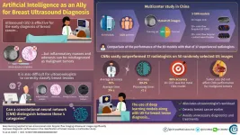
To tackle this limitation, Dr. He, in collaboration with scientists from 13 hospitals in China, conducted the largest multicenter study on breast ultrasound yet in an attempt to train convolutional neural networks (CNNs) to classify ultrasound images. As detailed in their paper published in Chinese Medical Journal, the scientists collected 15,648 images from 3,623 patients and used half of them to train and the other half to test three different CNN models. The first model only used 2D ultrasound intensity images as input, whereas the second model also included color flow Doppler images, which provide information on blood flow surrounding breast lesions. The third model further added pulsed wave Doppler images, which provide spectral information over a specific area within the lesions.
Each CNN consisted of two modules. The first one, the detection module, contained two main submodules whose overall task was to determine the position and size of the breast lesion in the original 2D ultrasound image. The second module, the classification module, received only the extracted portion from the ultrasound images containing the detected lesion. The output layer contained four categories corresponding to each of the four classifications of breast masses commonly used in China.
First, the scientists checked which of the three models performed better. The accuracies were similar and around 88%, but the second model including 2D images and color flow Doppler data performed slightly better than the other two. The reason the pulsed wave Doppler data did not contribute positively to performance may be that few pulsed wave images were available in the overall dataset. Then, researchers checked if differences in tumor size caused differences in performance. While larger lesions resulted in increased accuracy in benign tumors, size did not appear to have an effect on accuracy when detecting malignancies. Finally, the scientists put one of their CNN models to the test by comparing its performance to that of 37 experienced ultrasonologists using a set of 50 randomly selected images. The results were vastly in favor of the CNN in all regards, as Dr. He remarks: "The accuracy of the CNN model was 89.2%, with a processing time of less than two seconds. In contrast, the average accuracy of the ultrasonologists was 30%, with an average time of 314 seconds."
This study clearly showcases the capabilities of deep learning algorithms as complementary tools for the diagnosis of breast lesions through ultrasound. Moreover, unlike previous studies, the researchers included data obtained using ultrasound equipment from different manufacturers, which hints at the remarkable applicability of the trained CNN models regardless of the ultrasound devices present at each hospital. In the future, the integration of artificial intelligence into diagnostic procedures with ultrasound could speed up the early detection of cancer. It would also bring about other benefits, as Dr. He explains: "Because CNN models do not require any type of special equipment, their diagnostic recommendations could reduce predetermined biopsies, simplify the workload of ultrasonologists, and enable targeted and refined treatment."
Let us hope artificial intelligence soon finds a home in ultrasound image diagnostics so doctors can work smarter, not harder.
INFORMATION:
Reference
Titles of original papers: Deep learning applied to two-dimensional color Doppler flow imaging ultrasound images significantly improves diagnostic performance in the classification of breast masses: a multicenter study
Journal: Chinese Medical Journal
DOI: 10.1097/CM9.0000000000001329
X-Ray Experiments, Machine Learning Could Trim Years Off Battery R&D
By Glenn Roberts Jr.
An X-ray instrument at Berkeley Lab contributed to a battery study that used an innovative approach to machine learning to speed up the learning curve about a process that shortens the life of fast-charging lithium batteries.
Researchers used Berkeley Lab's Advanced Light Source, a synchrotron that produces light ranging from the infrared to X-rays for dozens of simultaneous experiments, to perform a chemical imaging technique known as scanning transmission ...
Parkinson's disease (PD) is well known as a debilitating disease that gradually worsens over time. Although the disease's progression has been largely tied to the loss of motor functions, non-motor symptoms, including the loss of cognitive abilities, often emerge early in the disease.
Much less understood is the role that specific neural circuits play in these distinct motor and non-motor functions.
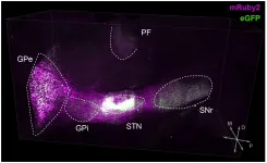
A new study led by neurobiologists at the University of California San Diego and their colleagues found that specific, identifiable neural pathways are charged with particular functions during stages of the disease. ...
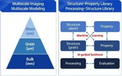
Developing new materials and novel processes has continued to change the world. The M3I3 Initiative at KAIST has led to new insights into advancing materials development by implementing breakthroughs in materials imaging that have created a paradigm shift in the discovery of materials. The Initiative features the multiscale modeling and imaging of structure and property relationships and materials hierarchies combined with the latest material-processing data.
The research team led by Professor Seungbum Hong analyzed the materials research projects reported by leading global ...
Researchers at the Bloomberg~Kimmel Institute for Cancer Immunotherapy at the Johns Hopkins Kimmel Cancer Center have developed DeepTCR, a software package that employs deep-learning algorithms to analyze T-cell receptor (TCR) sequencing data. T-cell receptors are found on the surface of immune T cells. These receptors bind to certain antigens, or proteins, found on abnormal cells, such as cancer cells and cells infected with a virus or bacteria, to guide the T cells to attack and destroy the affected cells.
"DeepTCR is an open-source software that ...
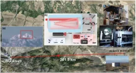
A research team led by Professor PAN Jianwei and Professor XU Feihu from University of Science and Technology of China achieved single-photon 3D imaging over 200 km using high-efficiency optical devices and a new noise-suppression technique, which is commented by the reviewer as an almost "heroic" attempt at single photon lidar imaging at very long distances.
Lidar imaging technology has enabled high precision 3D imaging of target scene in recent year. Single photon imaging lidar is an ideal technology for remote optical imaging with single-photon level sensitivity and picosecond resolution, yet its imaging range is strictly limited by the quadratically decreasing count of photons that echo back.
Researchers first optimized transceiver optics. The lidar system setup adopted ...
A new automated process prints a peptide-based hydrogel scaffold containing uniformly distributed cells. The scaffolds hold their shapes well and successfully facilitate cell growth that lasts for weeks.
"Bioprinting" -- 3D printing that incorporates living cells -- has the potential to revolutionize tissue engineering and regenerative medicine. Scientists have experimented with natural and synthetic "bioinks" to print out scaffolds that hold cells in place as they grow and form a tissue with a specific shape. But there are challenges with cell survival. Natural bioinks, such as gelatin and collagen, need to be treated with chemicals or ultraviolet light to hold their shape, which affects ...
A new analysis of the entire genetic makeup of more than 53,000 people offers a bonanza of valuable insights into heart, lung, blood and sleep disorders, paving the way for new and better ways to treat and prevent some of the most common causes of disability and death.
The analysis from the Trans-Omics for Precision Medicine (TOPMed) program examines the complete genomes of 53,831 people of diverse backgrounds on different continents. Most are from minority groups, which have been historically underrepresented in genetic studies. The increased representation should translate into better understanding of how heart, lung, blood and sleep disorders affect minorities and should help reduce longstanding health disparities.
"The Human Genome Project has generated ...
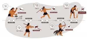
Researchers at Tel Aviv University were able to reconstruct the nutrition of stone age humans. In a paper published in the Yearbook of the American Physical Anthropology Association, Dr. Miki Ben-Dor and Prof. Ran Barkai of the Jacob M. Alkov Department of Archaeology at Tel Aviv University, together with Raphael Sirtoli of Portugal, show that humans were an apex predator for about two million years. Only the extinction of larger animals (megafauna) in various parts of the world, and the decline of animal food sources toward the end of the stone age, led humans to gradually increase the vegetable element in their nutrition, until finally they had no choice but to domesticate both plants and animals - and became farmers.
"So far, attempts to reconstruct the diet of stone-age humans ...
WASHINGTON, April 5, 2021 -- Forever chemicals are known for being water-, heat- and oil-resistant, which makes them useful in everything from rain jackets to firefighting foams. But the chemistry that makes them so useful also makes them stick around in the environment and in us -- and that could be a bad thing: https://youtu.be/tqKEG5LxPiY.
INFORMATION:
Reactions is a video series produced by the American Chemical Society and PBS Digital Studios. Subscribe to Reactions at http://bit.ly/ACSReactions and follow us on Twitter @ACSReactions.
The American Chemical Society (ACS) is a nonprofit organization chartered by the U.S. Congress. ACS' mission is to advance the broader ...
CORVALLIS, Ore. - Researchers in the Oregon State University College of Science have taken a key step toward new drugs and vaccines for combating COVID-19 with a deep dive into one protein's interactions with SARS-CoV-2 genetic material.
The virus' nucleocapsid protein, or N protein, is a prime target for disease-fighting interventions because of the critical jobs it performs for the novel coronavirus' infection cycle and because it mutates at a comparatively slow pace. Drugs and vaccines built around the work of the N protein carry the potential to be highly effective and for longer periods of time - i.e., less susceptible to resistance.
Among the SARS-CoV-2 proteins, ...
