Cambridge team uses powerful new MRI scans to enable life-changing surgery in first for adults with epilepsy
A new technique has enabled ultra-powerful magnetic resonance imaging (MRI) scanners to identify tiny differences in patients’ brains that cause treatment-resistant epilepsy. In the first study to use this approach, it has allowed doctors at Addenbrooke’s Hospital, Cambridge, to offer the patients surgery to cure their condition.
Previously, 7T MRI scanners – so called because they operate using a 7 Tesla magnetic field, more than double the strength of previous 3T scanners – have suffered from signal blackspots in crucial parts of the brain. But in research published today in Epilepsia, researchers in Cambridge and Paris have used a technique that overcomes this problem.
Around 360,000 people in the UK have a condition known as focal epilepsy, which causes seizures to spread from part of the brain. A third of these individuals have persistent seizures despite medication, and the only treatment that can cure their condition is surgery. Epileptic seizures are the sixth most common reason for hospital admission.
In order for surgeons to perform this operation, they need to be able to see the lesions (diseased tissue) in the brain responsible for the seizures. Then, they can work out exactly which areas to remove to cure the patient’s epilepsy. If surgeons are able to see the lesions on MRI scans, this can double the chances of the patient being free of seizures following surgery.
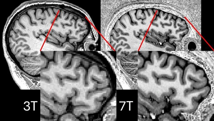
Ultra-high field 7T MRI scanners allow much more detailed resolution on brain scans and have been shown in other countries to be better than the NHS’s best 3T MRI scanners at detecting these lesions in patients with drug-resistant epilepsy (and in fact, most NHS hospitals have even weaker, 1.5T scanners). However, 7T MRI scans are susceptible to dark patches known as signal dropouts. These dropouts commonly occur in the temporal lobes, where most cases of epilepsy arise.
To overcome this problem, researchers at the University of Cambridge’s Wolfson Brain Imaging Centre, working with colleagues at the Université Paris-Saclay, trialled a technique known as ‘parallel transmit’, which uses eight transmitters around the brain rather than just one to avoid the problematic drop-outs.
Chris Rodgers, Professor of Biomedical Imaging at the University of Cambridge, said: “It used to be the case that MRI scanners used a single radio transmitter, but in a similar way to how single wifi routers leave areas where you will struggle to get a signal, so these scanners would tend to leave blackspots on brain scans where it was hard to make out the relevant tissue.
“Now, by using multiple radio transmitters positioned around the patients’ head – like having a wifi mesh around your home – we can get much clearer images with fewer blackspots. This is important for the epilepsy scans because we need to see very precisely which part of the brain is misbehaving.
“The Paris group’s plug-and-play sequences avoid the need to calibrate the scanner at every visit, making it practical to use these scans for scanning patients.”
The team tested their approach with 31 drug-resistant epilepsy patients recruited at Addenbrooke’s Hospital, part of Cambridge University Hospitals NHS Foundation Trust (CUH), to see if the parallel transmit 7T scanner was better than conventional 3T scanners at detecting brain lesions.
They found that the parallel transmit 7T scanner identified previously unseen structural lesions in nine patients. It confirmed in four patients suspected lesions detected using 3T scanners, and in a further four patients showed that suspected lesions could be disregarded.
Parallel transmit 7T images were clearer than conventional (‘single transmit’) 7T images in more than half of the cases (57%), and in the remaining cases the images were equally clear. Single transmit scanners never outperformed parallel transmit scanners.
As a result of their findings, more than half of the patients (18 patients, or 58%) had the management of their epilepsy changed. Nine patients were offered surgery to remove the lesion, and one patient was offered laser interstitial thermal therapy (which uses heat to remove the lesion). For three patients, scans showed more complex lesions, meaning that surgery was no longer an option. Five patients, because of the size or location of their lesions, were offered stereotactic electroencephalography (sEEG), a technique for pinpointing the lesions using electrodes inserted into the brain – this procedure is not used for everyone because it is very costly and invasive, and the 7T scans allowed it to be offered to the patients it was most likely to help.
Dr Thomas Cope from the University’s Department of Clinical Neurosciences, and a Consultant Neurologist at CUH, said: “Having epilepsy that doesn’t respond to anti-seizure medications can have a huge impact on patients’ lives, often affecting their independence and their ability to maintain a job. We know we can cure many of these patients, but that requires us to be able to pinpoint exactly where in the brain is the root of their seizures.
“7T scanners have shown promise over the past few years since their introduction, and now, thanks to this new technique, more epilepsy patients will be eligible for life-changing surgery.”
When the team asked patients about their experience afterwards, the patients reported only minor and occasional negative experiences, such as dizziness on scanner entry and additional claustrophobia from the head coil. This suggests that parallel transmit 7T MRI is acceptable to patients.
The research was supported by the Cambridge University Hospitals Academic Fund and the Medical Research Council, with support from the National Institute for Health and Care Research Cambridge Biomedical Research Centre.
Dr Cope is an Official Fellow at Murray Edwards College, Cambridge. Professor Rodgers is a Bye Fellow at Peterhouse, Cambridge.
“Once I’d had the surgery, it was very obviously the right decision”: Amanda Bradbury Amanda Bradbury, 29, wanted to be an interior designer when she was younger. She began a course at university, but despite it being a subject she really enjoyed, found herself overwhelmed, struggling to concentrate and increasingly anxious. Eventually it became too much, and she was forced to drop out.
What Amanda didn’t know was that her problems were being caused by a tiny flaw in her brain that was making her have seizures – so-called ‘focal epilepsy’.
At first, the most obvious signs of these seizures were auras, distortions in her vision. These began when she was around 19, but her symptoms started to become increasingly more frequent and problematic. She would often find herself getting extremely anxious, struggling to focus and follow conversations, forgetting things, having difficulty speaking or even swallowing.
“One of the things that would happen before a seizure is I'd get an intense feeling of fear, which I now realise was seizure-related.”
This began to have an impact on even the simplest things, she says. “I started leaving the house a lot less because of nerves, because it can affect your memory when you have a seizure. I'd be too nervous to talk, because I would get confused. I just got more and more unsure about what was going on.”
For some time, she dismissed her symptoms. But when she moved to Cambridge to live with her sister, it became harder to ignore what was happening.
“Because I was living with someone that knows me, [my sister] was able to see that certain things weren't making sense. I wasn't able to focus as much or I'd be saying words that didn’t relate to anything we were doing.”
Encouraged by her sister, she sought medical help. Doctors at Addenbrooke’s Hospital, Cambridge, diagnosed her as having focal epilepsy. Suddenly, it all made sense to her. But what was surprising was how frequently she was having these seizures. Although she thought they were occurring a couple of times a week, brainwave recordings revealed she was actually having them multiple times a day.
Amanda was given medication to manage her condition, but despite trying three different drugs, some of which at first appeared to reduce her symptoms, none of them were ultimately effective. This was when the doctors suggested surgery.
Amanda’s lesion was large enough to be visible to 3T MRI scanners (for many patients, the lesions are not clearly visible on these scanners, which is where ultra-high field 7T MRI scanners can help). The lesion was in her amygdala, the part of the brain responsible for controlling emotions, which explained why she would feel so fearful before and during an episode.
As the Cambridge team was able to pinpoint the lesion, surgeons could then remove it.
Very soon after surgery, Amanda began to feel different – less tired, with more energy and feeling less anxious. People around her noticed a difference, too, as she was able to focus and concentrate more.
She uses the example of an everyday task that to most of us would seem trivial to illustrate the difference surgery has made.
“One thing I can do a lot easier now is clean the kitchen!” she says. “I can stand up, focus on what I'm doing and be chatting while I'm doing it.”
She had become so used to having seizures and to struggling day to day that it took their disappearance to show her what normality was like. She now works in office administration, but wants to get back into interior design as a hobby.
“I want to try and do things that I enjoy, like interior design and things that are arty. I want to experience these things more.”
Although it took her some time to accept her diagnosis, describing it as “mentally overwhelming,” Amanda is very open about her epilepsy now. Becoming aware that she was a part a community of people living with the condition, that she was not going through these things alone, helped her, and so she wants to help others feel supported.
She also has no regrets about having had the surgery. Even though it felt like a big decision at the time, talking to the doctors and to her family made her realise that she was in safe hands and that this could really help her.
“Once I’d had the surgery, despite all the healing, it was very obviously the right decision,” she says. “Suddenly I realised I'm able to do loads of other things. It made me start to think, oh, what can I do? Things felt a lot more possible, like suddenly I'm able to do so many more things.”
Reference
Klodowski, K et al. Parallel transmit 7T MRI for adult epilepsy pre-surgical evaluation. Epilepsia; 21 March 2025: DOI: 10.1111/epi.18353
END
Previously, 7T MRI scanners – so called because they operate using a 7 Tesla magnetic field, more than double the strength of previous 3T scanners – have suffered from signal blackspots in crucial parts of the brain. But in research published today in Epilepsia, researchers in Cambridge and Paris have used a technique that overcomes this problem.
Around 360,000 people in the UK have a condition known as focal epilepsy, which causes seizures to spread from part of the brain. A third of these individuals have persistent seizures despite medication, and the only treatment that can cure their condition is surgery. Epileptic seizures are the sixth most common reason for hospital admission.
In order for surgeons to perform this operation, they need to be able to see the lesions (diseased tissue) in the brain responsible for the seizures. Then, they can work out exactly which areas to remove to cure the patient’s epilepsy. If surgeons are able to see the lesions on MRI scans, this can double the chances of the patient being free of seizures following surgery.
Ultra-high field 7T MRI scanners allow much more detailed resolution on brain scans and have been shown in other countries to be better than the NHS’s best 3T MRI scanners at detecting these lesions in patients with drug-resistant epilepsy (and in fact, most NHS hospitals have even weaker, 1.5T scanners). However, 7T MRI scans are susceptible to dark patches known as signal dropouts. These dropouts commonly occur in the temporal lobes, where most cases of epilepsy arise.
To overcome this problem, researchers at the University of Cambridge’s Wolfson Brain Imaging Centre, working with colleagues at the Université Paris-Saclay, trialled a technique known as ‘parallel transmit’, which uses eight transmitters around the brain rather than just one to avoid the problematic drop-outs.
Chris Rodgers, Professor of Biomedical Imaging at the University of Cambridge, said: “It used to be the case that MRI scanners used a single radio transmitter, but in a similar way to how single wifi routers leave areas where you will struggle to get a signal, so these scanners would tend to leave blackspots on brain scans where it was hard to make out the relevant tissue.
“Now, by using multiple radio transmitters positioned around the patients’ head – like having a wifi mesh around your home – we can get much clearer images with fewer blackspots. This is important for the epilepsy scans because we need to see very precisely which part of the brain is misbehaving.
“The Paris group’s plug-and-play sequences avoid the need to calibrate the scanner at every visit, making it practical to use these scans for scanning patients.”
The team tested their approach with 31 drug-resistant epilepsy patients recruited at Addenbrooke’s Hospital, part of Cambridge University Hospitals NHS Foundation Trust (CUH), to see if the parallel transmit 7T scanner was better than conventional 3T scanners at detecting brain lesions.
They found that the parallel transmit 7T scanner identified previously unseen structural lesions in nine patients. It confirmed in four patients suspected lesions detected using 3T scanners, and in a further four patients showed that suspected lesions could be disregarded.
Parallel transmit 7T images were clearer than conventional (‘single transmit’) 7T images in more than half of the cases (57%), and in the remaining cases the images were equally clear. Single transmit scanners never outperformed parallel transmit scanners.
As a result of their findings, more than half of the patients (18 patients, or 58%) had the management of their epilepsy changed. Nine patients were offered surgery to remove the lesion, and one patient was offered laser interstitial thermal therapy (which uses heat to remove the lesion). For three patients, scans showed more complex lesions, meaning that surgery was no longer an option. Five patients, because of the size or location of their lesions, were offered stereotactic electroencephalography (sEEG), a technique for pinpointing the lesions using electrodes inserted into the brain – this procedure is not used for everyone because it is very costly and invasive, and the 7T scans allowed it to be offered to the patients it was most likely to help.
Dr Thomas Cope from the University’s Department of Clinical Neurosciences, and a Consultant Neurologist at CUH, said: “Having epilepsy that doesn’t respond to anti-seizure medications can have a huge impact on patients’ lives, often affecting their independence and their ability to maintain a job. We know we can cure many of these patients, but that requires us to be able to pinpoint exactly where in the brain is the root of their seizures.
“7T scanners have shown promise over the past few years since their introduction, and now, thanks to this new technique, more epilepsy patients will be eligible for life-changing surgery.”
When the team asked patients about their experience afterwards, the patients reported only minor and occasional negative experiences, such as dizziness on scanner entry and additional claustrophobia from the head coil. This suggests that parallel transmit 7T MRI is acceptable to patients.
The research was supported by the Cambridge University Hospitals Academic Fund and the Medical Research Council, with support from the National Institute for Health and Care Research Cambridge Biomedical Research Centre.
Dr Cope is an Official Fellow at Murray Edwards College, Cambridge. Professor Rodgers is a Bye Fellow at Peterhouse, Cambridge.
“Once I’d had the surgery, it was very obviously the right decision”: Amanda Bradbury Amanda Bradbury, 29, wanted to be an interior designer when she was younger. She began a course at university, but despite it being a subject she really enjoyed, found herself overwhelmed, struggling to concentrate and increasingly anxious. Eventually it became too much, and she was forced to drop out.
What Amanda didn’t know was that her problems were being caused by a tiny flaw in her brain that was making her have seizures – so-called ‘focal epilepsy’.
At first, the most obvious signs of these seizures were auras, distortions in her vision. These began when she was around 19, but her symptoms started to become increasingly more frequent and problematic. She would often find herself getting extremely anxious, struggling to focus and follow conversations, forgetting things, having difficulty speaking or even swallowing.
“One of the things that would happen before a seizure is I'd get an intense feeling of fear, which I now realise was seizure-related.”
This began to have an impact on even the simplest things, she says. “I started leaving the house a lot less because of nerves, because it can affect your memory when you have a seizure. I'd be too nervous to talk, because I would get confused. I just got more and more unsure about what was going on.”
For some time, she dismissed her symptoms. But when she moved to Cambridge to live with her sister, it became harder to ignore what was happening.
“Because I was living with someone that knows me, [my sister] was able to see that certain things weren't making sense. I wasn't able to focus as much or I'd be saying words that didn’t relate to anything we were doing.”
Encouraged by her sister, she sought medical help. Doctors at Addenbrooke’s Hospital, Cambridge, diagnosed her as having focal epilepsy. Suddenly, it all made sense to her. But what was surprising was how frequently she was having these seizures. Although she thought they were occurring a couple of times a week, brainwave recordings revealed she was actually having them multiple times a day.
Amanda was given medication to manage her condition, but despite trying three different drugs, some of which at first appeared to reduce her symptoms, none of them were ultimately effective. This was when the doctors suggested surgery.
Amanda’s lesion was large enough to be visible to 3T MRI scanners (for many patients, the lesions are not clearly visible on these scanners, which is where ultra-high field 7T MRI scanners can help). The lesion was in her amygdala, the part of the brain responsible for controlling emotions, which explained why she would feel so fearful before and during an episode.
As the Cambridge team was able to pinpoint the lesion, surgeons could then remove it.
Very soon after surgery, Amanda began to feel different – less tired, with more energy and feeling less anxious. People around her noticed a difference, too, as she was able to focus and concentrate more.
She uses the example of an everyday task that to most of us would seem trivial to illustrate the difference surgery has made.
“One thing I can do a lot easier now is clean the kitchen!” she says. “I can stand up, focus on what I'm doing and be chatting while I'm doing it.”
She had become so used to having seizures and to struggling day to day that it took their disappearance to show her what normality was like. She now works in office administration, but wants to get back into interior design as a hobby.
“I want to try and do things that I enjoy, like interior design and things that are arty. I want to experience these things more.”
Although it took her some time to accept her diagnosis, describing it as “mentally overwhelming,” Amanda is very open about her epilepsy now. Becoming aware that she was a part a community of people living with the condition, that she was not going through these things alone, helped her, and so she wants to help others feel supported.
She also has no regrets about having had the surgery. Even though it felt like a big decision at the time, talking to the doctors and to her family made her realise that she was in safe hands and that this could really help her.
“Once I’d had the surgery, despite all the healing, it was very obviously the right decision,” she says. “Suddenly I realised I'm able to do loads of other things. It made me start to think, oh, what can I do? Things felt a lot more possible, like suddenly I'm able to do so many more things.”
Reference
Klodowski, K et al. Parallel transmit 7T MRI for adult epilepsy pre-surgical evaluation. Epilepsia; 21 March 2025: DOI: 10.1111/epi.18353
END