Scientists develop atomic force microscopy for imaging nanoscale dynamics of neurons
Researchers at the Max Planck Florida Institute for Neuroscience and Kanazawa University, Japan, have succeeded in imaging structural dynamics of living neurons with an unprecedented spatial resolution
2015-03-13
(Press-News.org) Atomic force microscopy (AFM) is a leading tool for imaging, measuring, and manipulating materials with atomic resolution - on the order of fractions of a nanometer.
AFM images surface topography of a structure by "touching" and "feeling" its surface by scanning an extremely fine needle (the diameter of the tip is about 5 nanometers, about 1/100 of light wavelength or 1/10,000 of a hair) on the surface.
This technique has been applied to image solid materials with nanometer resolution, but it has been difficult to apply AFM for a soft and large sample like eukaryotic cells and neurons without damaging the sample. Additionally, image acquisition with conventional AFM is too slow to capture fast cellular morphology changes.
Researchers have extensively modified the AFM system for imaging eukaryotic cells and neurons with high spatial and temporal resolution.
This new system allows for analysis of cell morphology changes with a spatial resolution ~20-100 fold better than that of a standard light microscope.
Imaging structural dynamics of living cells and neurons
While progress has been made over the past decades in the pursuit to optimize atomic force microscopy (AFM) for imaging living cells, there were still a number of limitations and technological issues that needed to be addressed before fundamental questions in cell biology could be address in living cells.
In their March publication in Scientific Reports, researchers at Max Planck Florida Institute for Neuroscience and Kanazawa University describe how they have built the new AFM system optimized for live-cell imaging. The system differs in many ways from a conventional AFM: it uses an extremely long and sharp needle attached to a highly flexible plate. The system is also optimized for fast scanning to capture dynamic cellular events. These modifications have enabled researchers to image living cells, such as mammalian cell lines or mature hippocampal neurons, without any sign of cellular damage.
"We've now demonstrated that our new AFM can directly visualize nanometer-scale morphological changes in living cells", explained Dr. Yasuda, neuroscientist and scientific director at the Max Planck Florida Institute for Neuroscience.
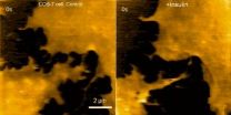
In particular, this study demonstrates the capability to track structural dynamics and remodeling of the cell surface, such as morphogenesis of filopodia, membrane ruffles, pit formation or endocytosis, in response to environmental stimulants. An example of this capability can be visualized in movie 1, where a fibroblast is imaged before and after treatment with insulin hormone, which intensely enhances the ruffling at the leading edge of the cell. Another example is seen in movie 2, where the morphological changes of a finger-like neuronal protrusion in the mature hippocampal neuron are observed.
According to Dr. Yasuda, the successful observations of structural dynamics in live neurons present the possibility of visualizing the morphology of synapses at nanometer resolution in real time in the near future. Since morphology changes of synapses underlie synaptic plasticity and our learning and memory, this will provide us with many new insights into mechanisms of how neurons store information in their morphology, how it changes synaptic strength and ultimately how it creates new memory.
INFORMATION:
Link to publication in Scientific Reports: http://www.nature.com/srep/2015/150304/srep08724/full/srep08724.html#supplementary-information
About Max Planck Florida Institute for Neuroscience
The Max Planck Florida Institute for Neuroscience (Jupiter, Florida, USA) specializes in the development and application of novel technologies for probing the structure, function and development of neural circuits. It is the first research institute of the Max Planck Society in the United States.
ELSE PRESS RELEASES FROM THIS DATE:
2015-03-13
Metastatic melanoma is the leading cause of skin cancer deaths in the United States; once melanoma has spread (metastasized), life expectancy for patients can be dramatically shortened. At present, the reference therapy for patients diagnosed with metastatic melanoma is Dacarbazine (DTIC), which is associated with poor patient outcomes.
In a study published in Molecular Cancer Research, March 12, 2015, the laboratory of Mary J.C. Hendrix, PhD, in collaboration with other scientists found that standard treatments for metastatic melanoma are not effective against a growth ...
2015-03-13
An FDA-approved drug for high blood pressure, guanabenz, prevents myelin loss and alleviates clinical symptoms of multiple sclerosis (MS) in animal models, according to a new study. The drug appears to enhance an innate cellular mechanism that protects myelin-producing cells against inflammatory stress. These findings point to promising avenues for the development of new therapeutics against MS, report scientists from the University of Chicago in Nature Communications on Mar. 13.
"Guanabenz appears to enhance the cell's own protective machinery to diminish the loss of ...
2015-03-13
Almost all patients with a group of blood cancers called B-cell malignancies have two prominent "fingerprints" on the surface of leukemia and lymphoma cancers, called CD22 and CD19, Vallera explained. To develop the drug, Vallera and colleagues chose two antibody fragments that each selectively bind to CD19 and CD22. They used genetic engineering to attach these two antibodies to a potent toxin, the bacterial diphtheria toxin. When the antibody fragments bind to the two targets on the cancer cell, the entire drug enters the cell, and the toxin kills the cell.
Vallera; ...
2015-03-13
Boston, Mass., USA - Today at the 93rd General Session and Exhibition of the International Association for Dental Research, researcher Bertha A. Chavez Gonzalez, Universidade de Minas Gerias, Lima, San Borja, Peru, will present a study titled "Birth Weight and Pregnancy Complications Associated With the Enamel Defects." The IADR General Session is being held in conjunction with the 44th Annual Meeting of the American Association for Dental Research and the 39th Annual Meeting of the Canadian Association for Dental Research.
This cross-sectional representative study ...
2015-03-13
Boston, Mass., USA - Today at the 93rd General Session and Exhibition of the International Association for Dental Research, researcher Aderonke A. Akinkugbe, University of North Carolina at Chapel Hill, USA, will present a study titled "Environmental Tobacco Smoke is Associated With Periodontitis in U.S. Non-smokers." The IADR General Session is being held in conjunction with the 44th Annual Meeting of the American Association for Dental Research and the 39th Annual Meeting of the Canadian Association for Dental Research.
Periodontitis affects approximately 47% of ...
2015-03-13
Leading public health researchers today [Friday 13 March 2015] call for the sale of tobacco to be phased out by 2040, showing that with sufficient political support and stronger evidence-based action against the tobacco industry, a tobacco-free world - where less than 5% of adults use tobacco - could be possible in less than three decades.
Writing in a major new Series in The Lancet, an international group of health and policy experts, led by Professors Robert Beaglehole and Ruth Bonita from the University of Auckland in New Zealand, call on the United Nations (UN) to ...
2015-03-13
Estoril, Portugal: Treating respiratory disease is often difficult because drugs have to cross biological barriers such as respiratory tissue and mucosa, and must therefore be given in large quantities in order for an effective amount to reach the target. Now researchers from Germany, Brazil and France have shown that the use of nanoparticles to carry antibiotics across biological barriers can be effective in treating lung infections. Doing so allows better delivery of the drug to the site of infection, and hence prevents the development of antibiotic resistance which ...
2015-03-12
Highlights
Among pregnant women, the risk for adverse pregnancy outcomes--such as preterm delivery or the need for neonatal intensive care--increased across stages of chronic kidney disease.
The risks of intrauterine death or fetal malformations were not higher in women with chronic kidney disease.
An estimated 26 million people in the United States have chronic kidney disease.
Washington, DC (March 12, 2015) -- Even mild kidney disease during pregnancy may increase certain risks in the mother and baby, according to a study appearing in an upcoming issue of ...
2015-03-12
BUFFALO, N.Y. - Political scientists at the University at Buffalo and Pennsylvania State University have published new research investigating how partisan differences in macroeconomic policy have contributed to substantial and rising economic inequality in the United States.
The negative consequences of such policy decisions, researchers found, have a greater impact on people at the lower end of the economic spectrum, but are "significantly more muted" for those at the higher end of the spectrum.
The study, "Partisan Differences in the Distributional Effects of Economic ...
2015-03-12
WORCESTER, MA - A new "app" for finding and mapping chromosomal loci using multicolored versions of CRISPR/Cas9, one of the hottest tools in biomedical research today, has been developed by scientists at the University of Massachusetts Medical School. This labeling system, details of which were published in PNAS and first presented at the American Society for Cell Biology/International Federation for Cell Biology annual meeting in Philadelphia in December, could be a key to understanding the spatial and temporal regulation of gene expression by allowing researchers to measure ...
LAST 30 PRESS RELEASES:
[Press-News.org] Scientists develop atomic force microscopy for imaging nanoscale dynamics of neurons
Researchers at the Max Planck Florida Institute for Neuroscience and Kanazawa University, Japan, have succeeded in imaging structural dynamics of living neurons with an unprecedented spatial resolution


