There and back again: how neurons make room for growth in a developing organ
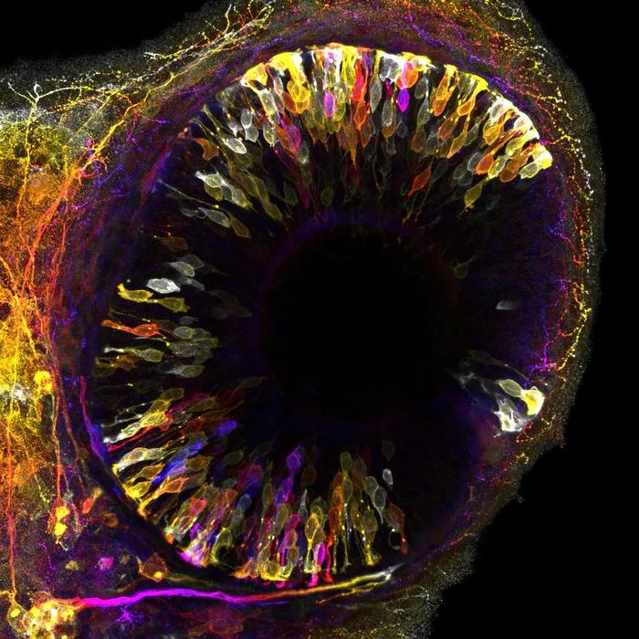
The researchers observed that an entire population of neurons, photoreceptors, temporarily relocates away from the zone of the tissue where they reside and must fulfill their function (Fig. 1). This active movement creates space for incoming progenitor cells that divide in this area and thereby produce more cells that latercontribute to the neuronal retina. Blockage of the movements of photoreceptors leads to congestion, forcing progenitor cells to divide in wrong place which in turn causes tissue malformation. Thus, by transiently moving away, neurons avoid interference with progenitor cells to ensure harmonious organ development.
To Mauricio Rocha-Martins, the first author of the study, “This is a curious migration phenomenon, in whichneurons move away just to then move back, ending up where they started. It highlights that neuronalmigration, as opposed to what was previously believed, does not only move neurons to their correct location but can also play a direct role in the coordination of organ development”.
The implications of this research extend beyond the field of retinal development. Simultaneous growth andacquisition of functional architecture is a hallmark of most developing organs; the new findings offer the possibilityto investigate whether other developing organs employ similar strategies. Moreover, it is known that defects in neuronal migration can cause severe brain malformations in humans. The findings that failed migration of neurons can have deleterious consequences beyond the positioning of neurons points to the importance of examining the interactions between cells to fully understand the causes of human developmental disorders.
The study was supported by the MPI-CBG, the FCG-IGC, the German Research Foundation (NO 1069/5-1), and an ERC Consolidator Grant (H2020 ERC-2018-CoG-81904).
END