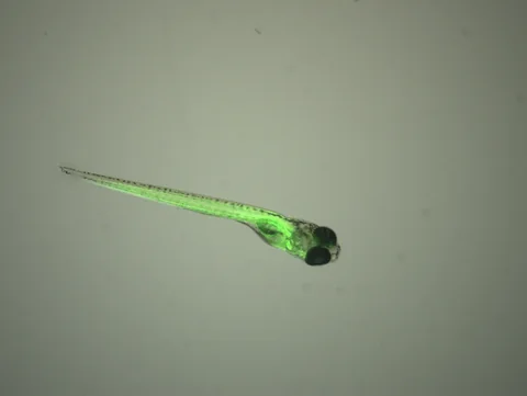
An organ rarely acts alone. Yet a microscope aimed at one tissue can miss what is happening elsewhere at the same moment. A Nature paper introduces a way to follow cellular activity across much of a small, transparent animal, making interactions between organs visible in a shared recording.
- The live signal reports changes in cellular calcium, rather than every process happening in a cell.
- The demonstrations use transparent fish; they are not whole-body cellular scans of people.
- Simultaneous signals suggest connections. Targeted experiments are needed to test causation.
- Publication
- Nature · 9 September 2026
- Platform
- WHOLISTIC
- Models
- Larval zebrafish; adult Danionella cerebrum proof of concept
- Readout
- Fluorescent calcium signals across tissues
Original publication: 9 Sep 2026 · The date above refers to this brief.
What the new method makes visible
Ruetten and colleagues combined fluorescent calcium sensors expressed widely across cells, volumetric microscopy and computational analysis. Their platform, WHOLISTIC, records changing signals across organs in larval zebrafish. They also demonstrated the approach in adult Danionella cerebrum, a small fish that remains transparent.
The study reports coordinated activity in structures including the kidney and digestive system, alongside interactions involving muscles and the nervous system. One line of experiments investigated how the brainstem helps redirect blood flow during low-oxygen exposure. These are studies of physiology in animal models, not tests of a therapy in patients.
Source 1 ↗A glowing cell is a measurement with a specific meaning
Calcium participates in many cellular processes. A sensor that changes fluorescence with calcium can reveal a pattern over time, but it does not provide a complete inventory of what that cell is doing. An apparently quiet trace is not proof that the cell is inactive.
The investigators also used expansion microscopy to examine structural and molecular context. That is a separate preparation-based approach to anatomy, not a claim that a living fish is enlarged while functioning normally. Keeping the live activity measurements separate from the structural maps helps readers understand what each image establishes.
Source 1 ↗From two signals moving together to a causal test
When two tissues change at similar times, at least three explanations deserve consideration: one influences the other, both respond to a third process, or the measurement couples them artificially. The paper combines recordings with targeted manipulations, including optogenetic experiments, for particular questions. That strengthens those specific tests; it does not turn every pair of synchronized traces into a proven circuit.
For your own reading notes, draw two arrows separately. One means “observed alongside.” The other means “changed after an intervention.” Then record exactly what was manipulated and what was measured. This simple distinction connects the work to our association-and-causation guide.
Source 1 ↗Why motion, transparency and analysis matter
A moving organ can make fluorescence appear to change even when the underlying signal does not. The platform therefore includes motion correction and methods for finding groups of signals with related timing. Those computational steps are part of the measurement, not just a way to make a prettier movie.
Transparent small animals also make optical access possible in ways that an opaque human body does not. The useful conclusion is that the method opens an experimental window in these models. Extending an insight to another species requires additional evidence about both the measurement and the underlying biology.
Source 1 ↗The next questions worth asking
Our proposed next step is to test a clearly specified interaction repeatedly: record two tissues, perturb one, measure the response in the other, and check whether a different intervention gives a consistent explanation. Independent laboratories could assess which patterns recur across animals and preparations.
A longer-term hope is to understand how a local disturbance spreads through a whole organism. That could generate hypotheses for disease research, but this paper does not establish a diagnostic tool or improved treatment. Better visibility is the beginning of an explanation; a reproducible causal account is the next task.
What each layer of evidence can tell you
| Layer | Useful information | Question still open |
|---|---|---|
| Live calcium recording | Where and when a measured signal changes | Which biological process explains the change? |
| Structural mapping | Which tissues and cells are nearby | Does anatomical proximity imply functional influence? |
| Coordinated timing | Which signals follow related patterns | Could another process drive both? |
| Targeted perturbation | What follows a specified intervention | Does the interpretation hold across controls and animals? |
Our reading framework, informed by the methods paper. These are different evidence types, not four measures of treatment effectiveness.
Your questions, answered
Can this technique scan every cell in a human body?
No. These demonstrations depend on optical access in small transparent fish and engineered fluorescent sensors. They do not establish a whole-body cellular imaging method for people.
Does a synchronized signal prove two organs communicate directly?
No. Shared timing can motivate a hypothesis. Testing a direct connection needs a design that addresses alternative explanations and the consequences of a targeted change.
Where does AI fit into the work?
Computational and machine-learning methods help extract and organize activity patterns. Those patterns still need biological interpretation and experimental checks; an algorithmically detected group is not, by itself, a confirmed mechanism.
What should I look for before calling this a medical breakthrough?
Specify the breakthrough: measurement, mechanism or patient benefit. Evidence that a method can reveal an animal’s physiology does not by itself establish either clinical usefulness or a safe treatment.
Limits of this interpretation
- Basic research in transparent animal models cannot establish benefit in patients.
- Calcium signals, optical access, sensor expression and motion handling constrain what is observed.
- The source check did not independently inspect raw recordings, code or supplementary movies.
Sources & transparency
- Ruetten, Zheng, Siwanowicz et al. (2026): Imaging cellular activity across all organs reveals body-wide circuits
Publisher HTML abstract, selected main-text results, methods and figure captions checked. Supplementary movies, raw data and code were not independently assessed. Original explanatory wording; no publisher figures are reproduced. The research article is CC BY 4.0. · Accessed 29 Sep 2026
DOI: 10.1038/s41586-026-10979-6 - Creative Commons Attribution 4.0 — licence for the Ruetten et al. article
Licence linked in the publisher’s rights statement. The explanation is newly written and includes our own reading framework and proposed next questions. · Accessed 29 Sep 2026
Prepared and source-checked with AI on 29 September 2026. Press-news Team is our collective publication byline, not a claim of medical credentials or human review. No human editorial or clinical review has taken place. This is educational reporting on research methods, not medical advice. Access limitations are listed with each source. Reading frameworks and proposed follow-up tests are our commentary, not additional study findings. The photograph is illustrative.
Source check: AI source check — selected primary research and reported numbers
Clinical review: Not applicable to this educational guide
Suggest a correction