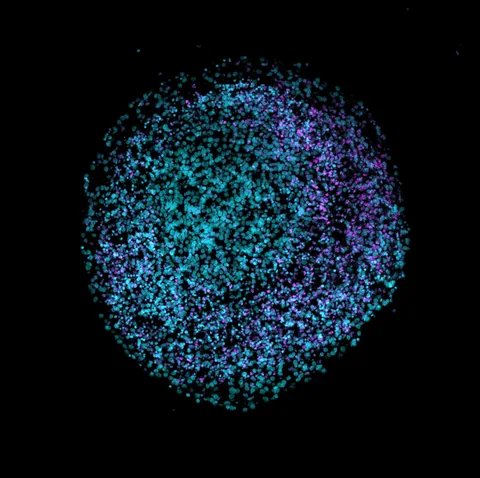
Lab-grown cortical organoids help scientists investigate developing neural tissue, but even structures grown side by side can differ. Researchers describe a way to track those differences before interpreting the biology.
- An organoid forming and an organoid showing the intended cortical features are distinct measurements.
- In an initial version of the format, six of 16 cell lines produced organoids meeting the authors’ described patterning criteria.
- A later quality score and manufacturing changes were used to refine comparisons; these laboratory models are not complete human brains.
- Paper date
- 3 November 2026
- Early comparison
- 16 cell lines
- Focus
- Formation versus patterning
Original publication: 3 Nov 2026 · The date above refers to this brief.
In the initial format, formation was not the only test
cell lines in the initial comparison
Follow the quality question
Form
Grow tissue-like structures from stem-cell lines.
Check
Measure whether the intended cortical features are present.
Compare
Interpret genetic differences in the selected samples and test their robustness.
Why make a miniature tissue model?
Cortical organoids are three-dimensional cell cultures that can reproduce selected features of early brain development. They let researchers compare cell behaviour under controlled laboratory conditions. Their resemblance to a developing tissue is partial: an organoid is not an adult human brain and does not capture every cell type or interaction.
Source 1 ↗The first number is not the whole outcome
The paper reports that its platform achieved approximately 100% organoid formation across 64 lines. Formation answers whether a structure was produced. It does not by itself establish that the resulting tissue has the intended cortical identity, cellular composition or useful consistency.
Source 1 ↗A different question gave a different denominator
In the early slit-well format, the authors examined 16 lines in eight isogenic sets. Six lines produced organoids that robustly displayed the described forebrain progenitor and cortical neuron markers at two months. By subtraction, ten did not meet that stated criterion. This is an early-format comparison, not the final success rate of the subsequently refined platform.
Source 1 ↗What did the quality checks add?
The researchers combined observations from different stages of growth with a transcriptomic quality index, CortiCOSI, to compare samples. The approach asks whether the measured signatures match the intended tissue model and whether unwanted cell populations are prominent. No single marker is presented as a complete definition of model quality.
Source 1 ↗Selection helps interpretation but needs transparency
The team applied quality-based selection to an existing dataset before comparing matched genetic lines. A cleaner comparison can reduce noise, while selection also changes which samples enter the analysis. Readers need to know which samples were excluded, why and whether the finding holds in additional lines and independent datasets.
Source 1 ↗Why it matters beyond organoids
The same issue appears in our guide to single-cell atlas settings: a label or quality threshold organises a dataset, but it also determines which observations are compared. The result is most useful when the choice and its limitations stay visible.
A formed model is not automatically a well-patterned model
Primary PDF: abstract, initial format comparison and patterning Results p. 2, and quality-control Results and Fig. 4. The initial-results page was visually inspected. Publisher metadata and abstract also checked. Supplements, source-data files, code and clinical records were not independently audited. No publisher figure reproduced.
01What was approximately 100%?
- What was observed
- The abstract reports approximately 100% organoid formation across 64 lines.
- Where the conclusion stops
- Formation is not the same endpoint as passing cortical patterning criteria.
Source 1 · Primary PDF abstract p. 1
02What did six of 16 measure?
- What was observed
- Six lines in the early format robustly produced organoids with the specified cortical markers.
- Where the conclusion stops
- This was before later refinement and is not the final platform success rate.
Source 1 · Primary PDF Results p. 2
Numbers you can inspect
| Measure | Value & unit | Origin & method |
|---|---|---|
| Reported formation dataset | 64 cell lines | Reported Lines in the abstract’s approximate formation figure, not people. Source 1 · Primary PDF abstract |
| Initial patterning comparison | 16 cell lines | Reported Lines in eight isogenic sets using the initial format. Source 1 · Primary PDF Results p. 2 |
| Lines meeting criteria | 6 cell lines | Reported Robust patterning under the stated marker criteria in the initial comparison. Source 1 · Primary PDF Results p. 2 |
| Other lines in comparison | 10 cell lines | Calculated 16 minus 6; not necessarily absent organoid formation. Source 1 · Primary PDF Results p. 2 |
Compare the actual experiments
These studies answer different questions. Read the unit and endpoint before comparing results.
| Study | Unit & setting | Readout | Interpretation boundary |
|---|---|---|---|
| Organoid formation Source 1 · Primary PDF abstract | Stem-cell lines | Whether a tissue-like structure formed | Formation does not establish cortical identity. |
| Early patterning assessment Source 1 · Primary PDF Results p. 2 | 16 lines in the initial format | Cortical marker criteria at two months | It is a different outcome and phase from the broader formation figure. |
The export includes claims, available numbers, methods and source locations. It contains our reading notes and published summaries; it is not raw participant data or an independent reanalysis.
Evidence update · 8 Nov 2026
First publication. Reported observations, arithmetic and proposed follow-up questions are separated. No raw-data reanalysis. AI source check; no human editorial or clinical review.
Formation and quality answer different questions
| Reading question | What the paper reports | What remains open |
|---|---|---|
| Formation | The authors report approximately 100% formation across 64 lines for the platform | A formed structure is not automatically well-patterned cortex |
| Initial patterning | Six of 16 lines met the described marker criteria in the early format | This is not the final success rate after refinement |
| Quality score | CortiCOSI combines selected transcriptomic indicators | A selected subset needs transparent reporting and replication |
| Clinical meaning | A laboratory model of selected neural features | It does not demonstrate a dementia treatment |
Original Press-News reading framework. Follow-up questions are our interpretation, not experiments performed by the authors.
Your questions, answered
Is a brain organoid a complete human brain?
No. It is a laboratory tissue model that reproduces selected features of early development.
Does almost 100% formation mean almost every organoid had the intended identity?
No. Formation and cortical patterning are different outcomes; the authors assessed quality separately.
What does six out of 16 refer to?
The number of lines meeting described cortical marker criteria in the early slit-well format, before later refinements.
Does this study show a treatment for dementia?
No. It studies the production and comparison of laboratory models, not treatment outcomes in patients.
Limits of this interpretation
- The six-of-16 figure describes an initial format and should not be presented as the final platform success rate.
- Cell lines, organoids and donors are different units; repeated structures do not automatically represent additional people.
- Quality-based selection can change the sample being compared and needs independent replication.
- The model lacks the full cellular and physiological context of a human brain; no treatment efficacy was tested.
Sources & transparency
- Integrated manufacturing platform for cortical organoids reveals early phosphatase and prefoldin dysregulation in MAPT V337M neurons
Primary PDF: abstract, initial format comparison and patterning Results p. 2, and quality-control Results and Fig. 4. The initial-results page was visually inspected. Publisher metadata and abstract also checked. Supplements, source-data files, code and clinical records were not independently audited. No publisher figure reproduced. · Accessed 8 Nov 2026
DOI: 10.1038/s44400-026-00147-y
Prepared and source-checked with AI. Press-news Team is the collective publication byline, not a medical reviewer. No human editorial or clinical review has taken place. This educational article examines research methods and laboratory or observational findings, not individual diagnosis or treatment. We did not conduct these experiments or reanalyse raw data. Reported results, our interpretation and proposed future tests are separated. Source-access limits are recorded below. Photographs are illustrative.
Source check: AI source check — selected primary PDF results, captions and study boundaries
Clinical review: Not applicable to this educational guide
Suggest a correction