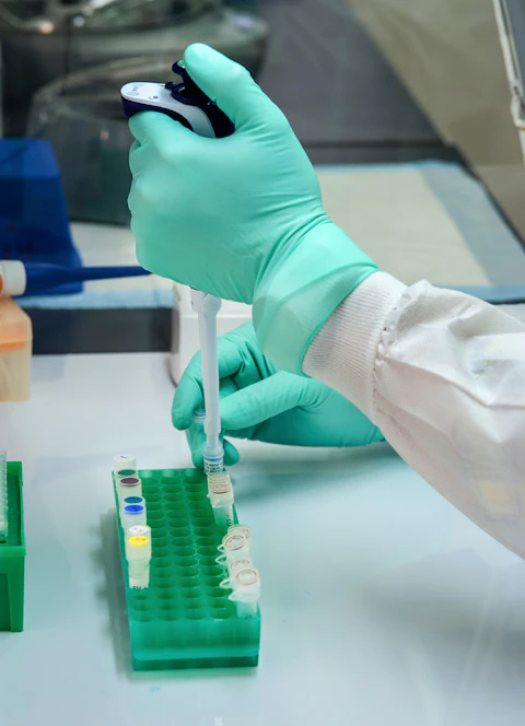
A broken DNA molecule can be joined or processed for another repair route. A new biochemical study shows why the temporary arrangement of its ends matters before the final repair reaction is complete.
- DNA-end synapsis means bringing ends together; ligation means sealing them.
- A catalytic mutant can help separate assembly from chemical activity.
- A purified yeast-protein system tests a mechanism, not repair success in a patient.
- Paper date
- 22 October 2026
- System
- Purified Saccharomyces cerevisiae proteins
- Key distinction
- A bridge versus a completed join
Original publication: 22 Oct 2026 · The date above refers to this brief.
Three questions for the repair claim
Assembly
Were the broken ends brought into a bridge?
Chemistry
Could the system seal the ends under that condition?
Processing
Did resection change when those outcomes were separated?
Two routes, one contested DNA end
Braunshier and colleagues reconstruct competition between non-homologous end joining, or NHEJ, and short-range DNA-end resection. Resection processes an end so that it can enter a recombination pathway. The authors find that a synapsed arrangement of broken ends strongly inhibits this processing in their purified system.
Source 1 ↗The control that makes the result useful
If joined DNA resisted processing, the explanation could simply be that the original broken substrate had disappeared. The study instead examines situations where the ends can be brought together without being successfully sealed. Non-ligatable ends and catalytically impaired ligase assemblies help separate those possibilities. Fig. 3 reports reduced resection even when ligation is inefficient or absent.
Source 1 ↗Assembly is an outcome in its own right
Our reading framework gives assembly and chemistry separate columns. Ask whether a perturbation prevents the molecular bridge, disables the final chemical reaction or does both. A mutant that changes both would be harder to interpret than one whose assembly behaviour is independently assessed.
This distinction also matters when reading an enzyme-inhibitor headline. Removing a protein and disabling its catalytic site can leave different structures behind. Neither manipulation is automatically equivalent to what a future drug would do in a living cell.
RPA changes a later part of the process
The paper also examines RPA, a single-stranded-DNA-binding protein. Under the tested conditions, it promotes removal of the end-bound Ku obstacle by stimulating MRX-associated exonuclease activity with phosphorylated Sae2. The authors distinguish this from stimulating the initial internal cut. That separation supports a more specific explanation than simply saying that RPA makes repair faster.
Source 1 ↗Keep the experiment attached to its replicate count
The Fig. 3 caption assigns three independent experiments to its ligation quantification and four to the resection quantification. Those are assay-specific denominators, not seven independent organisms or one shared sample size for the whole paper.
Source 1 ↗What would extend the mechanism?
Our proposed next question is whether changing bridge lifetime changes pathway use in a living system while preserving the other functions of the proteins. That would connect the purified mechanism to a different level of evidence; it is not an experiment we performed.
Read this beside our Cas9 article, which also separates an activated molecular state from successful chemical cutting. The analogy concerns experimental logic. The proteins, substrates and biological processes differ. This work helps explain a repair decision; it does not establish a cancer treatment or a way to improve an individual’s DNA repair.
A stopped reaction can still leave an active structure
Selected accepted primary PDF: initial pathway Results on p.2, end-joining/synapsis Results on p.4, Fig. 3 and RPA-related Results on p.6. Fig. 3 and caption visually inspected. Publisher HTML metadata and abstract also checked. Supplements, source-data files, code and clinical records were not independently audited. No publisher figure reproduced. The accepted version may receive further edits.
01Must DNA be sealed to resist resection?
- What was observed
- Synapsed assemblies with inefficient or disabled ligation still suppress resection.
- Where the conclusion stops
- The assembly itself needs verification; not every catalytic mutation preserves it.
Source 1 · PDF pp.4–6; Fig. 3
02Which RPA effect was resolved?
- What was observed
- Stimulation of downstream exonuclease processing and Ku removal rather than the initial incision.
- Where the conclusion stops
- This is a mechanistic distinction under the assay conditions, not overall repair efficacy.
Source 1 · PDF p.6; RPA Results
Numbers you can inspect
| Measure | Value & unit | Origin & method |
|---|---|---|
| Ligation quantification replicates | 3 independent experiments | Reported Fig. 3a caption denominator only. Source 1 · PDF p.6; Fig. 3a caption |
| Resection quantification replicates | 4 independent experiments | Reported Fig. 3c caption denominator only; not pooled with ligation. Source 1 · PDF p.6; Fig. 3c caption |
Compare the actual experiments
These studies answer different questions. Read the unit and endpoint before comparing results.
| Study | Unit & setting | Readout | Interpretation boundary |
|---|---|---|---|
| Catalytic ligase controls Source 1 · PDF pp.4–6; Fig. 3 | Purified yeast-protein/DNA assemblies | Ligation versus resection | Catalytic activity and bridge assembly are separable properties. |
| RPA processing assays Source 1 · PDF p.6; RPA Results | Ku-bound DNA ends and resection proteins | Downstream processing and obstacle removal | A specific biochemical step, not proof of better repair in vivo. |
The export includes claims, available numbers, methods and source locations. It contains our reading notes and published summaries; it is not raw participant data or an independent reanalysis.
Evidence update · 23 Oct 2026
First publication. Observations, our arithmetic and proposed follow-up questions are separated. No raw-data reanalysis. AI source check; no human editorial or clinical review.
Separate the stages before comparing the result
| Reading question | Evidence to retain | What remains open |
|---|---|---|
| Are the ends held together? | Synapsis measurement and relevant controls | How stable that assembly is in a cell |
| Are they chemically joined? | Ligation readout with the same protein state | Whether a failed reaction preserves the bridge |
| Are the ends processed? | Resection assay and its own normalisation | Which processing step changes |
| What is replicated? | Caption-specific independent experiments | Whether a count refers to assays, events or organisms |
Original Press-News assessment framework. Follow-up questions are our interpretation, not additional experiments performed by the authors.
Your questions, answered
Are synapsis and ligation the same thing?
No. Synapsis brings ends together; ligation seals them.
Why use a catalytically impaired ligase?
It helps ask whether the physical assembly can inhibit processing even when the joining reaction is disabled.
Did the paper study patients?
The mechanism discussed here comes from purified yeast proteins and associated biochemical and single-molecule experiments.
Does more resection always mean better repair?
No such general rule follows. The relevant endpoint is successful repair in context, not simply increasing one processing step.
Limits of this interpretation
- Reconstitution deliberately omits much of a living cell’s regulatory environment.
- Synapsis and ligation need distinct measurements and perturbation controls.
- Yeast-protein findings do not establish quantitative human repair outcomes.
- Raw traces, source-data archives and supplementary experiments were not independently audited.
Sources & transparency
- Braunshier, Cannavo, Morin et al. (2026): NHEJ-mediated DNA synapsis and RPA-driven MRX exonuclease balance DNA end resection to control DSB repair
Selected accepted primary PDF: initial pathway Results on p.2, end-joining/synapsis Results on p.4, Fig. 3 and RPA-related Results on p.6. Fig. 3 and caption visually inspected. Publisher HTML metadata and abstract also checked. Supplements, source-data files, code and clinical records were not independently audited. No publisher figure reproduced. The accepted version may receive further edits. · Accessed 23 Oct 2026
DOI: 10.1038/s41467-026-78670-y
Prepared and source-checked with AI. Press-news Team is the collective publication byline, not a medical reviewer. No human editorial or clinical review has taken place. This educational article examines research methods and laboratory findings, not individual diagnosis or treatment. We did not conduct these experiments or reanalyse raw data. Reported results, our interpretation and proposed future tests are separated. Source-access limits are recorded below. Photographs are illustrative.
Source check: AI source check — selected primary PDF results, captions and study boundaries
Clinical review: Not applicable to this educational guide
Suggest a correction