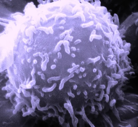
An immune cell can travel without responding well to the signals around it. A new laboratory study makes that distinction visible, then tests whether the physical properties of its culture medium can temporarily change the response.
- Migration, deformability and signal sensing are different outcomes.
- The seven-day priming experiment includes a washout test.
- Single-cell counts do not replace independent donor replication.
- Paper date
- 15 October 2026
- Material
- Activated human T cells outside the body
- Boundary
- Cell behaviour; no clinical rejuvenation endpoint
Original publication: 15 Oct 2026 · The date above refers to this brief.
The medium changed, not a measured percentage of immunity
culture-medium viscosity in centipoise (cP)
Keep the clock beside the effect
Condition
Record the physical setting and cell history.
Remove
Measure again after returning to baseline conditions.
Follow
Keep durability separate from the initial response.
The finding: a physical setting changes a cellular response
Dance and colleagues compare migration, deformation and sensing in T cells from younger and older donors. After seven days in an 8 cP medium, older cells showed improved movement towards a chemokine in microchannels. The response was particularly associated with memory CD4 cells. After transfer back to baseline medium, the seven-day-primed cells returned to baseline behaviour by day four.
Source 1 ↗Why the washout matters more than a rejuvenation headline
Our reading puts the time after removal beside the apparent improvement. A result measured immediately after conditioning answers whether cells can change; a washout experiment asks how long that change remains. Neither establishes durable protection from infection.
A useful future comparison would hold activation, donor and measurement timing constant while recording both the peak response and its decline. That would make two candidates with similar initial effects distinguishable. These are proposed evaluation questions, not a claim that the present study measured clinical protection.
A denominator check: cells are nested inside donors
The baseline migration analysis includes 15 younger and 28 older donors. Some mechanistic measurements use far fewer independent donors; for example, the membrane-tension analysis lists four. The authors also disclose a related patent application.
Source 1 ↗Build a donor-level result before counting more trajectories
Our suggested evidence sheet has one line per donor and separate columns for cell subset, activation history, conditioning duration and follow-up. A thousand tracked cells from one sample can describe that sample precisely while leaving uncertainty about other people.
This is also a useful connection to our subgroup guide: a response concentrated in a selected cell population should retain that population in the headline and in any attempted replication. Pooling subsets may hide both a responsive group and a group that did not change.
What would make the next step useful?
We would look for replication across additional independent donors, prespecified subset analyses and tests of functions beyond migration. The physical treatment and culture history should be stated explicitly so that a transient laboratory response is reproducible.
The future opportunity is a controllable experimental handle on cell behaviour. Whether it improves meaningful immune function, persists after transfer or behaves safely in a living organism requires separate evidence. This work does not support changing diet, blood viscosity or any treatment. Read it beside the yeast-confinement study: both make the physical setting an experimental variable, using different materials and endpoints.
Behaviour, duration and independent replication
Selected accepted primary PDF: baseline migration Results and Fig. 1 on pp.2–3; priming and washout on pp.9–11; Fig. 6 caption on p.12. Fig. 5 caption visually inspected. Accepted version may receive edits. Publisher HTML metadata and abstract also checked. Supplements, source-data files, code and clinical records were not audited. No publisher figure reproduced.
01What improved?
- What was observed
- Selected surveillance behaviours after conditioning in elevated-viscosity medium.
- Where the conclusion stops
- Not a clinical endpoint or whole-body age reversal.
Source 1 · Accepted PDF pp.9–11; Fig. 5
02Does the seven-day effect remain?
- What was observed
- Behaviour returns to baseline by day four after washout in that experiment.
- Where the conclusion stops
- Longer priming schedules are separate experiments.
Source 1 · Accepted PDF pp.9–10
Numbers you can inspect
| Measure | Value & unit | Origin & method |
|---|---|---|
| Control viscosity | 0.77 cP | Reported Reported culture condition, not an efficacy measure. Source 1 · PDF pp.9–11; Fig. 5 |
| Elevated viscosity | 8 cP | Reported Reported culture condition. Source 1 · PDF pp.9–11; Fig. 5 |
| Priming duration in washout comparison | 7 days | Reported Do not confuse conditioning duration with time after removal. Source 1 · PDF pp.9–11 |
| Baseline migration younger donors | 15 donors | Reported Baseline migration sample, not each downstream experiment. Source 1 · Fig. 1 caption, PDF p.3 |
| Baseline migration older donors | 28 donors | Reported Independent donor count for baseline migration. Source 1 · Fig. 1 caption, PDF p.3 |
| Membrane-tension donors | 4 donors | Reported Two younger and two older; single-cell counts are a different denominator. Source 1 · Fig. 6g caption, PDF p.12 |
Compare the actual experiments
These studies answer different questions. Read the unit and endpoint before comparing results.
| Study | Unit & setting | Readout | Interpretation boundary |
|---|---|---|---|
| Baseline migration Source 1 · PDF pp.2–3; Fig. 1 | Human donor-derived cells | Tracked movement | Migration alone does not test every surveillance feature. |
| Conditioning and washout Source 1 · PDF pp.9–11; Fig. 5 | Activated T cells in microchannels | Response after removal from elevated viscosity | Transient ex vivo behaviour, not clinical immune protection. |
The export includes claims, available numbers, methods and source locations. It contains our reading notes and published summaries; it is not raw participant data or an independent reanalysis.
Evidence update · 19 Oct 2026
First publication. Reported observations, our calculations and proposed follow-up questions are distinguished. No raw data reanalysis. AI source check; no human editorial or clinical review.
From movement to immune function: which claim does each test answer?
| Observation | Keep beside it | Next useful question |
|---|---|---|
| Cells move | Trajectory and donor identity | Does the cell respond to a relevant signal? |
| A response improves | Subset and activation history | Does it recur across independent donors? |
| A change persists | Time since conditioning ended | What remains after a prespecified washout? |
| Immune function improves | A functional endpoint beyond migration | Does a new study test that endpoint directly? |
Original Press-News comparison framework. Proposed checks are our interpretation, not additional experiments reported by the authors.
Your questions, answered
Were older people given a viscosity treatment?
No. The work examined cultured human T cells outside the body.
Does young-like mean the immune system became young?
No. It describes selected cell behaviours in the specified laboratory assays.
Why do donor numbers matter?
Measurements from the same donor share a biological source; they are not independent people.
What is the useful next question?
Whether a reproducible change in a defined cell subset persists and improves a separately measured function.
Limits of this interpretation
- Activated cells ex vivo do not reproduce an intact immune system.
- Donor replication differs between the baseline survey and mechanistic assays.
- The displayed seven-day priming effect is transient after washout.
- Supplementary files and raw trajectories were not independently audited.
Sources & transparency
- Dance, Amitrano, Macaluso et al. (2026): Acute priming using elevated fluid viscosity recovers young-like single-cell surveillance behaviors in aged human T cells
Selected accepted primary PDF: baseline migration Results and Fig. 1 on pp.2–3; priming and washout on pp.9–11; Fig. 6 caption on p.12. Fig. 5 caption visually inspected. Accepted version may receive edits. Publisher HTML metadata and abstract also checked. Supplements, source-data files, code and clinical records were not audited. No publisher figure reproduced. · Accessed 19 Oct 2026
DOI: 10.1038/s41467-026-78411-1
Prepared and source-checked with AI. Press-news Team is a collective publication byline, not a medical reviewer. No human editorial or clinical review has taken place. This educational explainer examines experimental methods and basic or preclinical research; it does not provide individual diagnosis or treatment advice. We did not conduct these experiments or reanalyse raw data. Findings, interpretation and proposed future tests are separated. Source-access limits are recorded below. Photographs are illustrative.
Source check: AI source check — selected primary PDF results, captions and experimental boundaries
Clinical review: Not applicable to this educational guide
Suggest a correction