After injury, cells in the intestinal lining can change their behaviour. Researchers used several models to study a population that appears during repair of the mouse colon.
- The main experiments investigate a cell population that emerges after irradiation in a mouse colon model.
- Lineage tracing, gene-expression measurements and human organoids provide complementary but different evidence.
- A human organoid is tissue grown in the laboratory; the study did not test a treatment for patients.
- Paper date
- 17 November 2026
- Core models
- Mouse colon and organoids
- Question
- Cell states during repair
Original publication: 17 Nov 2026 · The date above refers to this brief.
Reading an experimental repair story
Observe
Identify a changing cell population after injury in a mouse model.
Trace
Examine marked cells and related molecular signals in separate assays.
Compare
Look for related signatures in human organoids without claiming a patient treatment.
A lining that can change
The cells lining the intestine are continually renewed. The authors studied what happens when the mouse colon is injured by irradiation and identified a population marked by expression of a gene called Ysc. Their experiments suggest that some of these cells can move toward a stem-like state during repair.
Source 1 ↗A map and a lineage answer different questions
One gene-expression analysis grouped 11,722 mouse epithelial cells into 11 categories. The authors also used lineage tracing to examine what happened to cells carrying a selected marker. A cell-state map indicates which molecular signatures are present; lineage tracing provides different evidence about the descendants of marked cells.
Source 1 ↗Fields of view are not mice
For one Figure 2 quantification, the paper reports six microscopic fields and three mice per group. Another Figure 2 analysis used ten fields with three mice per group. Fields, cells and animals are different sampling units and should not be presented as independent patients.
Source 1 ↗What was observed in human organoids?
The team also examined human colonic and small-intestinal organoids grown outside the body. In one gene-expression comparison the figure labels 8,101 cells from the non-irradiated condition and 8,761 from the irradiated condition. These are cell counts in a laboratory analysis, not participant counts or proof that the same repair process works in patients.
Source 1 ↗A proposed molecular pathway
The authors report experiments involving p53, YSC and YAP, and propose a pathway connecting the observed cell state to regeneration in their models. The strength of the conclusion differs by model: manipulating a pathway in mice and observing a related molecular signature in organoids do not answer identical questions.
Source 1 ↗What comes next?
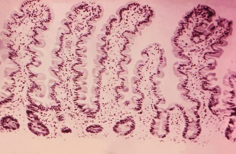
Further work could test the proposed mechanism in independent organoid preparations and additional injury models. It would also be necessary to establish relevance and safety in people before drawing conclusions about therapy. The accompanying CDC photograph shows normal small-intestinal tissue, not the mouse colon or an organoid from this study.
A mouse-cell map and human organoid comparison are not patient counts
Primary PDF: abstract, Results, Fig. 2 caption p. 5 and Fig. 6 caption p. 10. Both caption pages were visually inspected. Publisher metadata and abstract also checked. Supplements, source-data files, code and clinical records were not independently audited. No publisher figure reproduced.
01What does 11,722 count?
- What was observed
- The paper reports 11,722 mouse epithelial cells in the cell-state analysis.
- Where the conclusion stops
- Cells are not independent patients or the number of animals.
Source 1 · Primary PDF Results and Fig. 1 pp. 2, 4
02Did the human analysis involve treated patients?
- What was observed
- Figure 6 compares cells from non-irradiated and irradiated human organoids in the laboratory.
- Where the conclusion stops
- Organoids are not whole people and do not establish clinical treatment benefit.
Source 1 · Primary PDF Fig. 6 caption p. 10
Numbers you can inspect
| Measure | Value & unit | Origin & method |
|---|---|---|
| Mouse epithelial cells mapped | 11722 cells | Reported Cells in the mouse epithelial-state analysis. Source 1 · Primary PDF Results p. 2 |
| Mouse cell categories | 11 groups | Reported Cell groups in the reported epithelial analysis. Source 1 · Primary PDF Results p. 2 |
| Microscopy fields in one quantification | 6 fields | Reported Fields in Figure 2E; from three mice per group. Source 1 · Primary PDF Fig. 2 caption p. 5 |
| Mice per group for that quantification | 3 mice | Reported Independent animal count in the selected figure panel. Source 1 · Primary PDF Fig. 2 caption p. 5 |
| Non-irradiated organoid cells | 8101 cells | Reported Cell count in the selected human-organoid expression comparison. Source 1 · Primary PDF Fig. 6 caption p. 10 |
| Irradiated organoid cells | 8761 cells | Reported Cell count in the selected human-organoid expression comparison. Source 1 · Primary PDF Fig. 6 caption p. 10 |
Compare the actual experiments
These studies answer different questions. Read the unit and endpoint before comparing results.
| Study | Unit & setting | Readout | Interpretation boundary |
|---|---|---|---|
| Mouse cell-state and lineage tracing Source 1 · Primary PDF Results and Fig. 2 | Cells, image fields and mice | Gene-expression states and marked-cell descendants | Sampling units differ across panels. |
| Human intestinal organoids Source 1 · Primary PDF Fig. 6 caption p. 10 | Lab-grown organoid cells | Expression signatures in two conditions | A laboratory model does not establish a patient outcome. |
The export includes claims, available numbers, methods and source locations. It contains our reading notes and published summaries; it is not raw participant data or an independent reanalysis.
Evidence update · 17 Nov 2026
First publication. Reported observations, arithmetic and proposed follow-up questions are separated. No raw-data reanalysis. AI source check; no human editorial or clinical review.
Three models, three kinds of evidence
| Reading question | What the paper reports | What remains open |
|---|---|---|
| Mouse epithelial map | 11,722 cells grouped by gene expression | Cells are not individual mice |
| Mouse lineage tracing | Six image fields from three mice per group in one panel | Fields are nested within animals |
| Human organoids | 8,101 and 8,761 cells in two conditions | Lab-grown cells are not patient outcomes |
| Proposed pathway | p53, YSC and YAP examined in the models | Clinical relevance requires separate work |
Original Press-News reading framework. Follow-up questions are our interpretation, not experiments performed by the authors.
Your questions, answered
Were 11,722 patients treated?
No. The number refers to mouse epithelial cells in a gene-expression analysis.
Are six microscopic fields six independent mice?
No. One figure reports six fields from three mice per group.
Did the human organoids prove a clinical treatment works?
No. They are laboratory-grown tissue models, not treated patients.
Is the image the actual mouse colon from this experiment?
No. It is a separate CDC micrograph of normal human small intestine.
Limits of this interpretation
- Most mechanistic experiments use a specific irradiation injury model in mice.
- Human intestinal organoids are laboratory systems, not a patient treatment trial.
- Cells, microscopic fields and animals are different units of analysis.
- Cell-state signatures can support a hypothesis without proving that all human tissues behave the same way.
Sources & transparency
- Zhang, Zang, Song et al. (2026): The p53-YSC-YAP axis mediates enterocyte dedifferentiation during colonic regeneration after irradiation
Primary PDF: abstract, Results, Fig. 2 caption p. 5 and Fig. 6 caption p. 10. Both caption pages were visually inspected. Publisher metadata and abstract also checked. Supplements, source-data files, code and clinical records were not independently audited. No publisher figure reproduced. · Accessed 17 Nov 2026
DOI: 10.1038/s41421-026-00935-z
Prepared and source-checked with AI. Press-news Team is the collective publication byline, not a medical reviewer. No human editorial or clinical review has taken place. This educational article examines research methods and laboratory or observational findings, not individual diagnosis or treatment. We did not conduct these experiments or reanalyse raw data. Reported results, our interpretation and proposed future tests are separated. Source-access limits are recorded below. Photographs are illustrative.
Source check: AI source check — selected primary PDF results, captions and study boundaries
Clinical review: Not applicable to this educational guide
Suggest a correction