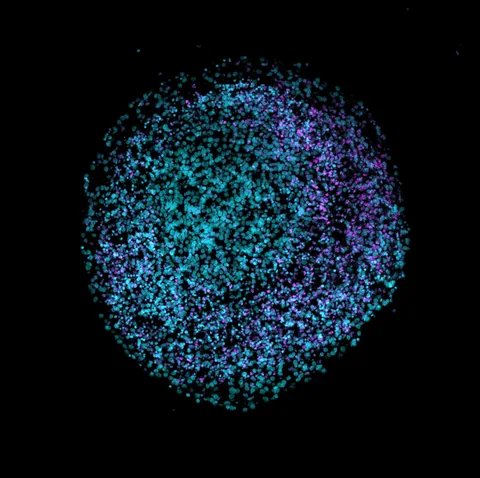
A thin image slice can hide the shape of a network. Researchers captured three-dimensional movies at selected stages as stem cells formed two kinds of organoid.
- Human stem-cell-derived lung and intestinal organoids were studied as laboratory models, not complete organs or patients.
- The researchers reported stage-prediction accuracy of 65% for lung and 74% for intestine in their respective test sets.
- Test and training sets were separated by organoid, but many organoids came from the same differentiation, limiting claims about independent replication.
- Paper date
- 16 November 2026
- Models
- Lung and intestinal organoids
- Prediction test
- Held-out organoids
Original publication: 16 Nov 2026 · The date above refers to this brief.
How the data were split
percent of single-cell data
From organoid to prediction
Image
Capture cell and mitochondrial structure in three dimensions over time.
Measure
Extract selected shape and movement features from single cells.
Test
Predict developmental stage on held-out organoids and describe the remaining dependence on differentiation.
Why add a fourth dimension?
Mitochondria form connected structures that change shape and move inside cells. The authors used lattice light-sheet microscopy to capture three spatial dimensions over time, then used computational tools to describe individual cells and their mitochondrial networks. A two-dimensional slice can miss connections that extend above or below the image plane.
Source 1 ↗What was actually grown?
The study images human stem cells and organoids at selected stages along paths toward branching lung and intestinal organoid models. The paper does not claim to follow each individual organoid continuously through the entire differentiation. Organoids reproduce some features of developing tissues under laboratory conditions; they do not reproduce every cell, blood supply or function of a complete human organ. The illustrative image on this page is a brain organoid from a different project.
Source 1 ↗Two dimensions can change the interpretation
In one comparison, the authors report that 3D analysis found changes in mitochondrial branching that the corresponding 2D analysis did not reveal. This is a comparison of analyses in the tested samples, not proof that all previous 2D work is wrong. Image quality and computational segmentation can also affect what is measured.
Source 1 ↗What did the predictions measure?
A random-forest model used measured cellular and mitochondrial features to classify developmental stages. The paper reports 65% accuracy for the lung model and 74% for the intestinal model. These are different classification problems: the paper describes five lung stages and four intestinal stages, so the numbers should not be treated as a simple contest between organs.
Source 1 ↗A held-out organoid is not a new laboratory
The authors split data by organoid, placing approximately 74% of single-cell data in training and 26% in testing. This avoids putting cells from the same organoid in both sets. However, many organoids in a condition came from one differentiation, so the result mainly estimates performance on held-out organoids within that differentiation, not on independently repeated differentiations.
Source 1 ↗What would strengthen the finding?
Repeating the workflow with independent differentiations, cell lines and laboratories would help test how well the patterns generalize. The paper also notes that measured movement includes active and passive motion that its current data do not distinguish. Our earlier organoid article explains why forming an organoid and establishing its quality are different questions.
A held-out organoid is not an independent differentiation
Accepted primary PDF: abstract, Fig. 1, Results and Fig. 7 p. 9, and Discussion. The Fig. 7 page was visually inspected; the accepted version may be edited. Publisher metadata and abstract also checked. Supplements, source-data files, code and clinical records were not independently audited. No publisher figure reproduced.
01Was the model tested on the same organoid used for training?
- What was observed
- The authors report splitting the data by organoid, with about 26% of single-cell data held out.
- Where the conclusion stops
- Many organoids per condition came from one differentiation, limiting claims of broader replication.
Source 1 · Accepted PDF Results p. 8 and Methods pp. 13–14
02Do 65% and 74% measure the same classification problem?
- What was observed
- The paper reports lung and intestinal stage accuracies in separate tasks.
- Where the conclusion stops
- There are five lung stages and four intestinal stages; neither number is patient diagnostic accuracy.
Source 1 · Accepted PDF Fig. 7 p. 9
Numbers you can inspect
| Measure | Value & unit | Origin & method |
|---|---|---|
| Training data | 74 percent of single-cell data | Reported Reported training share; split by organoid. Source 1 · Accepted PDF Results p. 8 |
| Held-out test data | 26 percent of single-cell data | Reported Reported test share; split by organoid. Source 1 · Accepted PDF Results and Methods pp. 8, 13–14 |
| Lung stage accuracy | 65 percent | Reported Reported accuracy for the lung-stage classification task. Source 1 · Accepted PDF Fig. 7 p. 9 |
| Intestinal stage accuracy | 74 percent | Reported Reported accuracy for a separate intestinal-stage classification task. Source 1 · Accepted PDF Fig. 7 p. 9 |
Compare the actual experiments
These studies answer different questions. Read the unit and endpoint before comparing results.
| Study | Unit & setting | Readout | Interpretation boundary |
|---|---|---|---|
| 2D and 3D image analyses Source 1 · Accepted PDF Fig. 1 p. 3 | Mitochondrial networks in selected cells | Branching patterns | A slice and a volume can yield different observations. |
| Held-out-organoid prediction Source 1 · Accepted PDF Results and Fig. 7 pp. 8–9 | Single-cell features from organoids | Developmental-stage classification | Held-out organoids can still share a differentiation. |
The export includes claims, available numbers, methods and source locations. It contains our reading notes and published summaries; it is not raw participant data or an independent reanalysis.
Evidence update · 17 Nov 2026
First publication. Reported observations, arithmetic and proposed follow-up questions are separated. No raw-data reanalysis. AI source check; no human editorial or clinical review.
What a validation result does and does not establish
| Reading question | What the paper reports | What remains open |
|---|---|---|
| 3D plus time | Network shape and movement tracked in laboratory cells | Not direct imaging of a patient |
| Lung-stage classifier | 65% accuracy in the reported test | Five stages; not a clinical score |
| Intestinal-stage classifier | 74% accuracy in the reported test | Four stages; not directly comparable to the lung task |
| Split by organoid | Cells from one organoid were kept in one split | Independent differentiations were not the main test |
Original Press-News reading framework. Follow-up questions are our interpretation, not experiments performed by the authors.
Your questions, answered
Did researchers scan patients?
No. The work studies human stem-cell-derived organoids in the laboratory.
Does 74% mean the intestinal model is clinically accurate?
No. It is developmental-stage classification accuracy for the study’s intestinal model, not a clinical test.
Were the test organoids independent of all training experiments?
The split was by organoid, but many organoids in a condition came from the same differentiation.
Is the image on this page from this study?
No. It shows a brain organoid from a separate project, as stated in the image credit.
Limits of this interpretation
- Organoids are laboratory models, not complete organs or clinical tests.
- The two classification tasks have different numbers of developmental stages.
- Held-out organoids may share a differentiation with training data; independent differentiations remain important.
- The accepted manuscript may change; motion cannot be fully separated into active and passive components in these data.
Sources & transparency
- McMahon, Agarwal, Arora et al. (2026): 4D mitochondrial network assumes distinct and predictive phenotypes through human lung and intestinal epithelial development
Accepted primary PDF: abstract, Fig. 1, Results and Fig. 7 p. 9, and Discussion. The Fig. 7 page was visually inspected; the accepted version may be edited. Publisher metadata and abstract also checked. Supplements, source-data files, code and clinical records were not independently audited. No publisher figure reproduced. · Accessed 17 Nov 2026
DOI: 10.1038/s41467-026-78762-9
Prepared and source-checked with AI. Press-news Team is the collective publication byline, not a medical reviewer. No human editorial or clinical review has taken place. This educational article examines research methods and laboratory or observational findings, not individual diagnosis or treatment. We did not conduct these experiments or reanalyse raw data. Reported results, our interpretation and proposed future tests are separated. Source-access limits are recorded below. Photographs are illustrative.
Source check: AI source check — selected primary PDF results, captions and study boundaries
Clinical review: Not applicable to this educational guide
Suggest a correction