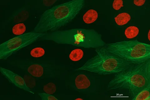
A brighter image is useful only if we know what it resolves. Researchers combined two-photon illumination with an oblique imaging plane to follow molecular signals in several experimental models.
- The paper reports approximately 300 nm lateral and 650 nm axial resolution, plus a separate localization precision below 60 nm in vivo.
- The reported speed comparison is with two-photon point scanning; contrast and resolving-power comparisons use linear excitation.
- The models include epithelial tissue, stem-cell-derived gastruloids and fruit-fly embryos, not a clinical diagnostic study.
- Paper date
- 13 November 2026
- Lateral / axial resolution
- About 300 / 650 nm
- Models
- Tissue, gastruloids and fly embryos
Original publication: 13 Nov 2026 · The date above refers to this brief.
Reported spatial resolution by direction
nanometres (approximate)
From light to a biological interpretation
Illuminate
Use a thin oblique excitation plane in the experimental sample.
Measure
Separate resolution, localization, contrast and imaging rate.
Interpret
Ask whether the demonstrated sample and comparison match the question.
Why depth changes the picture
In a complex sample, scattered light and differences in refractive index can make background fluorescence harder to separate from the signal. The authors describe a microscope that uses two-photon excitation and an oblique imaging plane while collecting light through a single primary objective. Their aim is to observe fine molecular activity in three-dimensional samples, not to test patients.
Source 1 ↗Resolution is not localization precision
The paper reports about 300 nanometres of lateral resolution and 650 nanometres axially. These describe the instrument’s ability to distinguish nearby structure in different directions under the reported conditions. Its reported localization precision below 60 nanometres in vivo is a different quantity: estimating where a detectable signal is, rather than separating every pair of objects at that distance. The values should not be presented as interchangeable.
Source 1 ↗Faster than which reference?
The authors report more than five times faster volumetric acquisition than their two-photon point-scanning comparison, reaching up to 3.25 million volume elements per second. That is an imaging rate in a stated comparison, not a count of cells, patients or diagnostic accuracy. It should not be generalized to every imaging task or microscope.
Source 1 ↗A separate comparison looks at contrast
In multicellular settings, the paper also reports about twice the contrast across the illumination depth and more than twice the resolving power relative to linear excitation. These comparisons answer a different question from the speed comparison. Improvements depend on the samples and settings used, and the paper does not establish that every specimen receives the same benefit.
Source 1 ↗What did the authors actually image?
Examples include epithelial tissue, stem-cell-derived gastruloids and living fruit-fly embryos. The work reports molecular measurements including transcription-factor dynamics and single-mRNA tracking. A gastruloid is a research model of aspects of early development; it is not a complete embryo or a clinical test.
Source 1 ↗What would the next comparison need?
Independent comparisons across more specimen types, laboratories and imaging conditions would clarify where the benefit holds and what trade-offs remain. The authors disclose a provisional patent application and commercial interests involving some contributors. Our earlier explanation of live-cell chemical imaging examines another balance between speed, signal and spatial detail.
Resolution, precision and speed answer different questions
Accepted primary PDF: abstract, Results, Fig. 1 p. 3 and Fig. 2. The Fig. 1 page was visually inspected; the accepted version may be edited. Publisher metadata and abstract also checked. Supplements, source-data files, code and clinical records were not independently audited. No publisher figure reproduced.
01Does the sub-60 nm number describe spatial resolution?
- What was observed
- The abstract labels it localization precision; it reports approximately 300 nm lateral and 650 nm axial spatial resolution.
- Where the conclusion stops
- Different measurement concepts; neither number establishes clinical accuracy.
Source 1 · Accepted PDF abstract p. 1 and Fig. 2
02What is the stated speed comparison?
- What was observed
- The paper reports more than five times faster volumetric acquisition compared with two-photon point scanning.
- Where the conclusion stops
- This is not the same comparator used for the linear-excitation contrast claim.
Source 1 · Accepted PDF abstract p. 1
Numbers you can inspect
| Measure | Value & unit | Origin & method |
|---|---|---|
| Approximate lateral resolution | 300 nm | Reported Reported lateral spatial resolution. Source 1 · Accepted PDF abstract p. 1 |
| Approximate axial resolution | 650 nm | Reported Reported axial spatial resolution. Source 1 · Accepted PDF abstract p. 1 |
| Localization precision threshold | 60 nm | Reported Reported as less than 60 nm in vivo; a distinct measurement. Source 1 · Accepted PDF abstract p. 1 |
| Maximum reported voxel rate | 3250000 voxels per second | Reported Up to this rate under the stated conditions. Source 1 · Accepted PDF abstract p. 1 |
Compare the actual experiments
These studies answer different questions. Read the unit and endpoint before comparing results.
| Study | Unit & setting | Readout | Interpretation boundary |
|---|---|---|---|
| Two-photon point-scanning comparison Source 1 · Accepted PDF abstract p. 1 | Volume elements per second | Reported acquisition speed | It does not measure clinical accuracy. |
| Linear-excitation comparison Source 1 · Accepted PDF abstract and Fig. 3 | Complex multicellular samples | Contrast and resolving power | A separate comparison from imaging speed. |
The export includes claims, available numbers, methods and source locations. It contains our reading notes and published summaries; it is not raw participant data or an independent reanalysis.
Evidence update · 16 Nov 2026
First publication. Reported observations, arithmetic and proposed follow-up questions are separated. No raw-data reanalysis. AI source check; no human editorial or clinical review.
Four numbers that do not mean the same thing
| Reading question | What the paper reports | What remains open |
|---|---|---|
| Lateral resolution | About 300 nm | A reported spatial resolution, not a localization error |
| Axial resolution | About 650 nm | A measurement in a different direction |
| Localization precision | Below 60 nm in vivo | A separate estimate, not the distance between two resolvable objects |
| Imaging rate | Up to 3.25 million voxels per second | Depends on the imaging setup and comparison |
Original Press-News reading framework. Follow-up questions are our interpretation, not experiments performed by the authors.
Your questions, answered
Does below 60 nm mean the microscope resolves every pair of structures 60 nm apart?
No. The reported localization precision and spatial resolution are different measures.
Was the microscope evaluated as a medical diagnostic test?
No. The reported demonstrations use research models, including tissue, gastruloids and fly embryos.
What is the speed compared with?
The paper compares volumetric acquisition speed with two-photon point scanning under the reported conditions.
Are there disclosed commercial interests?
Yes. The paper reports a provisional patent application and commercial interests involving some authors.
Limits of this interpretation
- The quantitative figures refer to the instrument and comparisons described in the accepted paper, not a universal guarantee.
- Resolution, localization precision, contrast and voxel rate are different measures.
- The study uses experimental models and does not evaluate a patient-facing diagnostic test.
- The accepted article may change; authors disclose a patent application and commercial interests.
Sources & transparency
- Keomanee-Dizon, Clenman, Duran et al. (2026): Depth-enhanced molecular imaging with two-photon oblique plane microscopy
Accepted primary PDF: abstract, Results, Fig. 1 p. 3 and Fig. 2. The Fig. 1 page was visually inspected; the accepted version may be edited. Publisher metadata and abstract also checked. Supplements, source-data files, code and clinical records were not independently audited. No publisher figure reproduced. · Accessed 16 Nov 2026
DOI: 10.1038/s41467-026-78668-6
Prepared and source-checked with AI. Press-news Team is the collective publication byline, not a medical reviewer. No human editorial or clinical review has taken place. This educational article examines research methods and laboratory or observational findings, not individual diagnosis or treatment. We did not conduct these experiments or reanalyse raw data. Reported results, our interpretation and proposed future tests are separated. Source-access limits are recorded below. Photographs are illustrative.
Source check: AI source check — selected primary PDF results, captions and study boundaries
Clinical review: Not applicable to this educational guide
Suggest a correction