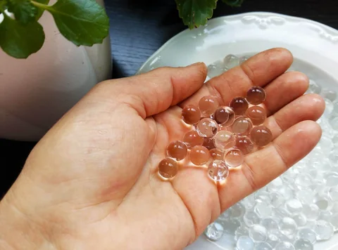
The inside of a cell is crowded, but it is not still. To study how a chemical reaction might change movement in a crowded environment, researchers built a much simpler model outside a living cell.
- The authors studied protein-rich droplets and compared active and inactive enzyme conditions.
- Fluorescence recovery and nanoparticle tracking measure related but distinct aspects of movement.
- A simplified droplet can test a mechanism without reproducing the whole cell.
- Paper date
- 30 October 2026
- Material
- Model protein-rich droplets
- Focus
- Enzyme activity and molecular mobility
Original publication: 30 Oct 2026 · The date above refers to this brief.
Model-derived protein diffusion in two droplet conditions
square micrometres per second
From activity to an interpretation
Build
Compare defined protein-rich model droplets.
Measure
Observe recovery, activity and tracer movement separately.
Interpret
Ask which conclusions depend on the physical model.
The question behind the droplets
Proteins and other molecules can concentrate into regions called biomolecular condensates. Dindo and colleagues use a simplified protein-rich droplet system to ask how crowding and enzyme activity influence one another. The model helps isolate physical effects that are difficult to disentangle inside a living cell.
Source 1 ↗How recovery becomes a measurement
In fluorescence recovery after photobleaching, researchers dim a small labelled region and observe fluorescence returning as molecules move. A shorter recovery time can indicate faster movement under the model and experimental conditions. The recovery curve is an observation; a diffusion coefficient derived from it also depends on the analysis model.
Source 1 ↗Two values, one model
The authors report model-derived BSA diffusion coefficients of 0.0092 ± 0.0001 and 0.015 ± 0.005 square micrometres per second in inactive and active droplets, respectively. The uncertainty bands in Fig. 3 arise from the conversion used for the fluorescence-recovery analysis. Our chart displays the central estimates only; the uncertainty belongs with those estimates.
Source 1 ↗Do not mix the sample sizes
For the Fig. 2b fluorescence-recovery conditions, the paper reports 12–23 independent droplets from different preparations. Its enzymatic-activity measurements were performed separately in three biologically independent experiments. Those numbers count different experimental units; they should not be combined into 36–69 independent replications.
Source 1 ↗A second way to follow movement
The team also tracked nanoparticles in active and inactive droplets. Particle trajectories offer a different readout from the recovery of labelled BSA, so agreement between them is informative, but the particle and protein diffusion values should not be treated as interchangeable. Simulations provide a further mechanistic interpretation rather than a direct view of every molecular interaction.
Source 1 ↗From a model back to biology
A reasonable next question is whether similar relationships hold in specific living-cell condensates with their own proteins, chemistry and boundaries. That is a proposed follow-up, not a result. Our Nemuri article describes a different laboratory system involving bacterial ribosome organisation; the shared language of condensates does not prove the same mechanism operates in both studies.
The experimental unit changes with the assay
Selected published primary PDF: model system and fluorescence-recovery Results on pp. 2–4, Fig. 2 caption on p. 4 and diffusion model on pp. 3–5. Fig. 2 page was visually inspected. Publisher HTML metadata and abstract also checked. Supplements, source-data files, code and clinical records were not independently audited. No publisher figure reproduced.
01What is compared in the chart?
- What was observed
- Two model-derived diffusion estimates for labelled BSA in inactive and active droplets.
- Where the conclusion stops
- They are central estimates for a simplified system; uncertainty bands matter.
Source 1 · Published PDF pp. 3–5; Fig. 3
02What do the sample counts mean?
- What was observed
- Fig. 2b reports 12–23 independent droplets and three separate biological enzyme-activity experiments.
- Where the conclusion stops
- Droplets and enzyme-activity experiments are not the same unit.
Source 1 · Published PDF p. 4; Fig. 2 caption
Numbers you can inspect
| Measure | Value & unit | Origin & method |
|---|---|---|
| Inactive-droplet BSA diffusion | 0.0092 square micrometres per second | Reported Central model-derived estimate; reported uncertainty ±0.0001. Source 1 · PDF pp. 3–5; Fig. 3 |
| Active-droplet BSA diffusion | 0.015 square micrometres per second | Reported Central model-derived estimate; reported uncertainty ±0.005. Source 1 · PDF pp. 3–5; Fig. 3 |
| Minimum Fig. 2b droplet count | 12 independent droplets | Reported Lower end of the 12–23 range per fluorescence-recovery condition. Source 1 · PDF p. 4; Fig. 2 caption |
| Maximum Fig. 2b droplet count | 23 independent droplets | Reported Upper end of the range per fluorescence-recovery condition. Source 1 · PDF p. 4; Fig. 2 caption |
| Enzyme-activity experiments | 3 biologically independent experiments | Reported A separate assay from the fluorescence-recovery measurements. Source 1 · PDF p. 4; Fig. 2 caption |
Compare the actual experiments
These studies answer different questions. Read the unit and endpoint before comparing results.
| Study | Unit & setting | Readout | Interpretation boundary |
|---|---|---|---|
| Fluorescence recovery Source 1 · PDF pp. 3–5 | Labelled BSA in individual model droplets | Recovery curve and model-derived diffusion | Depends on the model used to infer diffusion. |
| Tracer tracking Source 1 · PDF pp. 4–5; Fig. 3 | Nanoparticles in active and inactive droplets | Particle trajectories and derived motion | A tracer is not the same molecular species as BSA. |
The export includes claims, available numbers, methods and source locations. It contains our reading notes and published summaries; it is not raw participant data or an independent reanalysis.
Evidence update · 31 Oct 2026
First publication. Reported observations, arithmetic and proposed follow-up questions are separated. No raw-data reanalysis. AI source check; no human editorial or clinical review.
What each measurement can tell you
| Reading question | Evidence to retain | What remains open |
|---|---|---|
| Fluorescence recovery | How labelled protein signal returns after a region is bleached | A direct measurement of every molecule in a living cell |
| Diffusion model | A coefficient inferred from a recovery curve and model | A model-free measurement of cellular health |
| Nanoparticle tracks | Motion of tracer particles in selected droplets | The same diffusion value as the labelled protein |
| Enzyme assay | Reaction activity measured separately | A count of independently imaged droplets |
Original Press-News reading framework. Follow-up questions are our interpretation, not experiments performed by the authors.
Your questions, answered
Were these droplets taken from patients?
No. They are a simplified laboratory model assembled for this experiment.
Are twelve droplets the same as twelve independent enzyme experiments?
No. The figure caption describes droplet-based recovery measurements and separate biological enzyme-activity experiments.
Does faster recovery prove every molecule moves faster?
No. Recovery is measured for the labelled signal and interpreted with a model; other molecules may behave differently.
Could the work help explain cellular condensates?
It suggests questions for living-cell experiments, but the model alone cannot establish that the same quantitative effect occurs in cells.
Limits of this interpretation
- These engineered droplets isolate selected features of crowding and do not reproduce an intact cell.
- Model-derived diffusion coefficients and directly tracked nanoparticle motion are different readouts.
- Different panels use different enzymes, conditions and experimental units.
- We did not independently audit raw microscopy, code or supplementary analyses.
Sources & transparency
- Dindo, Metson, Ren et al. (2026): Enzymes can activate and mobilize dense biomolecular condensates across scales
Selected published primary PDF: model system and fluorescence-recovery Results on pp. 2–4, Fig. 2 caption on p. 4 and diffusion model on pp. 3–5. Fig. 2 page was visually inspected. Publisher HTML metadata and abstract also checked. Supplements, source-data files, code and clinical records were not independently audited. No publisher figure reproduced. · Accessed 31 Oct 2026
DOI: 10.1038/s41467-026-77666-y
Prepared and source-checked with AI. Press-news Team is the collective publication byline, not a medical reviewer. No human editorial or clinical review has taken place. This educational article examines research methods and laboratory or observational findings, not individual diagnosis or treatment. We did not conduct these experiments or reanalyse raw data. Reported results, our interpretation and proposed future tests are separated. Source-access limits are recorded below. Photographs are illustrative.
Source check: AI source check — selected primary PDF results, captions and study boundaries
Clinical review: Not applicable to this educational guide
Suggest a correction