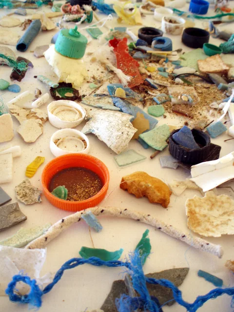
Immune cells help clear material from the lungs. A set of laboratory experiments asks how manufactured plastic particles interact with those cells and their surrounding tissue.
- The study used manufactured polystyrene particles, human-derived cells and lung slices, and mice under experimental conditions.
- The Methods describe blood from six de-identified donors for preparing monocyte-derived macrophages; this does not mean every experiment involved six donors.
- The findings do not quantify a person’s risk from everyday environmental exposure.
- Paper date
- 9 November 2026
- Models
- Human-derived tissue and cells; mice
- Material
- Manufactured polystyrene particles
Original publication: 9 Nov 2026 · The date above refers to this brief.
Three levels of evidence
Expose
Use manufactured particles in defined laboratory models.
Measure
Record cellular, tissue and animal-model readouts.
Translate
Ask separately whether and how findings apply to real human exposures.
What did the team put to the test?
The authors studied polystyrene particles in several settings: human monocyte-derived macrophages, cell lines, human precision-cut lung slices and mice. Macrophages are immune cells that can take up particles. Each model answers a different question; the results should not be pooled as if they came from one clinical trial.
Source 1 ↗A closer look at one cell measurement
In one laboratory experiment using human monocyte-derived macrophages, the paper reports an average of 48 particles per cell at day one and 46.5 at day six. Figure 1B also shows an intermediate time point. These are reported averages in a particular culture experiment, not a measurement of particles in a person’s lungs or proof of a steady decline over time.
Source 1 ↗Different readouts, different conclusions
Other experiments measured indicators of cell viability, metabolism and immune-cell activity, while mouse experiments examined tissue and cellular responses. The authors report changes in several of these readouts. A result in a cell line, a slice or a mouse can motivate further work, but it is not the same as demonstrating a disease outcome in people.
Source 1 ↗Six donors is not six replications of everything
The Methods describe material from six de-identified blood donors used to prepare macrophages. Different panels and models have their own units and sample sizes. The donor count should not be attached automatically to every figure or treated as the number of people whose health was followed.
Source 1 ↗Where translation remains uncertain
The particles were manufactured polystyrene, and the experiments used defined laboratory exposures. Environmental microplastics vary in composition, size and exposure history. This paper does not establish a safe threshold, a population disease rate or the effects of a specific everyday exposure.
Source 1 ↗A separate view from the field
Our companion wildlife article describes detections in selected tapirs and anteaters and associations with their surroundings. Those measurements involve different species, samples and particles. Reading the papers together is useful for generating questions, but neither links an individual wild animal’s blood result to the laboratory cell responses.
A laboratory result is not a personal exposure estimate
Accepted primary PDF: abstract, Methods on donor-derived cells, Fig. 1 and Results on pp. 4–6. The Fig. 1 and Results page was visually inspected; the accepted version may be edited. Publisher metadata and abstract also checked. Supplements, source-data files, code and clinical records were not independently audited. No publisher figure reproduced.
01What does the six-person count describe?
- What was observed
- The Methods describe blood from six de-identified donors for preparing macrophages.
- Where the conclusion stops
- It should not be assumed to be the denominator of every experiment or a clinical cohort.
Source 1 · Accepted PDF Methods p. 3
02Were the day-one and day-six numbers patient measurements?
- What was observed
- One cultured-cell experiment reports averages of 48 and 46.5 particles per macrophage at days one and six.
- Where the conclusion stops
- These are laboratory cell averages, with an intermediate point also shown, not personal lung counts.
Source 1 · Accepted PDF Fig. 1B and Results p. 5
Numbers you can inspect
| Measure | Value & unit | Origin & method |
|---|---|---|
| Blood donors for macrophage preparation | 6 donors | Reported Methods count; not necessarily the denominator of each panel. Source 1 · Accepted PDF Methods p. 3 |
| Reported average at day one | 48 particles per macrophage | Reported Reported cultured-cell average in one experiment. Source 1 · Accepted PDF Results p. 5 |
| Reported average at day six | 46.5 particles per macrophage | Reported Reported cultured-cell average; an intermediate time point is also shown. Source 1 · Accepted PDF Fig. 1B and Results p. 5 |
Compare the actual experiments
These studies answer different questions. Read the unit and endpoint before comparing results.
| Study | Unit & setting | Readout | Interpretation boundary |
|---|---|---|---|
| Human-derived macrophage culture Source 1 · Accepted PDF Methods and Fig. 1 | Cultured immune cells | Particle uptake and cellular readouts | Cell averages are not patient outcomes. |
| Mouse experiment Source 1 · Accepted PDF abstract and Results | Experimentally exposed mice | Tissue and cellular responses | A separate model does not directly quantify human environmental risk. |
The export includes claims, available numbers, methods and source locations. It contains our reading notes and published summaries; it is not raw participant data or an independent reanalysis.
Evidence update · 13 Nov 2026
First publication. Reported observations, arithmetic and proposed follow-up questions are separated. No raw-data reanalysis. AI source check; no human editorial or clinical review.
What each model contributes
| Reading question | What the paper reports | What remains open |
|---|---|---|
| Human-derived macrophages | Particle uptake and cellular readouts in culture | Not a direct measure of disease risk |
| Human lung slices | Responses of tissue maintained outside the body | Not a complete living respiratory system |
| Mice | Selected whole-animal experimental responses | Not a direct human exposure estimate |
| Environmental exposure | Different particles and real-world conditions remain to be studied | No safe or unsafe personal threshold was established |
Original Press-News reading framework. Follow-up questions are our interpretation, not experiments performed by the authors.
Your questions, answered
Did the study measure everyday health risk in people?
No. The experiments do not quantify personal risk from everyday exposure.
Were human cells used?
Yes. The authors describe human monocyte-derived macrophages and human lung slices, studied outside the body.
Does six donors mean six people were clinically followed?
No. The Methods describe blood from six de-identified donors for preparing macrophages; the count is not a clinical follow-up cohort.
Were all environmental plastics tested?
No. The study used selected manufactured polystyrene particles under experimental conditions.
Limits of this interpretation
- The exposure materials and conditions were experimental and do not reproduce every environmental exposure.
- Human-derived cells and lung slices were studied outside the body; the mouse model is a separate system.
- Six blood donors are described for macrophage preparation, not necessarily every analysis or a clinical follow-up.
- The accepted paper may be edited; we did not independently audit raw data, supplements or clinical records.
Sources & transparency
- Dhupar, Udoh, Noureen et al. (2026): Microplastics inhibit macrophage bioenergetics impairing homeostatic function and immune responsiveness
Accepted primary PDF: abstract, Methods on donor-derived cells, Fig. 1 and Results on pp. 4–6. The Fig. 1 and Results page was visually inspected; the accepted version may be edited. Publisher metadata and abstract also checked. Supplements, source-data files, code and clinical records were not independently audited. No publisher figure reproduced. · Accessed 13 Nov 2026
DOI: 10.1038/s41598-026-76299-x
Prepared and source-checked with AI. Press-news Team is the collective publication byline, not a medical reviewer. No human editorial or clinical review has taken place. This educational article examines research methods and laboratory or observational findings, not individual diagnosis or treatment. We did not conduct these experiments or reanalyse raw data. Reported results, our interpretation and proposed future tests are separated. Source-access limits are recorded below. Photographs are illustrative.
Source check: AI source check — selected primary PDF results, captions and study boundaries
Clinical review: Not applicable to this educational guide
Suggest a correction