When a cell moves across a surface, the trail it leaves can contain more than a footprint. A new laboratory study describes membrane-bound structures associated with the rear edge of migrating cancer cells.
- The authors describe migration-associated vesicles approximately 2–5 micrometres across in their tested systems.
- DNA analysis of vesicle material is not the same as sequencing every individual vesicle.
- The work is a laboratory characterisation, not a validated test for cancer spread.
- Paper date
- 28 October 2026
- Material
- Laboratory cell models and vesicle preparations
- Focus
- Formation, size and cargo
Original publication: 28 Oct 2026 · The date above refers to this brief.
Separate the cell, the preparation and the claim
Observe
Follow vesicle formation in a defined cell model.
Characterise
Measure the size and cargo in the tested material.
Validate
Ask what separate evidence is needed in another setting.
What are MoVes?
Tsering and colleagues call the structures motile vesicles, or MoVes. In the systems highlighted by the paper, they arise as migrating cancer cells retract their trailing edge. Microscopy and additional molecular analyses were used to characterise how the structures form and what they contain.
Source 1 ↗A size range, not a universal cut-off
The authors report MoVes of roughly 2–5 micrometres. For scale, multiplying by 1,000 converts this to approximately 2,000–5,000 nanometres. That is a reported range in the tested preparations, not a size threshold that can identify every extracellular vesicle in every sample.
Source 1 ↗What the DNA experiment examined
The paper reports genomic DNA spanning the parent-cell genome in MoVes material and compares genetic features of a cancer cell line with vesicle-associated DNA. An analysis of a vesicle preparation can establish what is present in that material; it does not by itself prove that every individual vesicle contains a complete genome.
Source 1 ↗A comparison is not a perfect classifier
The authors report vesicle formation in multiple cancer-cell lines and rare observations in the non-cancer cell types they examined. The frequency depends on the sampled cells, migration conditions and methods. A result described as rare in selected controls is not evidence that a future test would perfectly distinguish cancer from every non-cancer state.
Source 1 ↗Observation versus mechanism
The paper uses live imaging, electron microscopy and molecular assays to examine formation and contents. The authors also discuss possible functional consequences and future work. A plausible route from a laboratory observation to tumour progression is a research question that needs additional testing, not an outcome established for a particular person.
Why the image matters
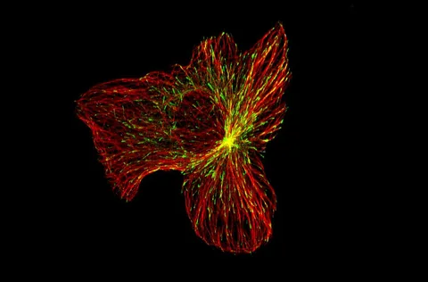
The illustrative fluorescence image accompanying this article shows a different migrating U2OS cancer cell, not a MoVe from this study. Our guide to cell-division structures makes a related distinction: the method, specimen and time point matter when interpreting what an image actually shows.
A pooled preparation and an individual vesicle are not the same unit
Selected published primary PDF: abstract and Results on pp. 1–2, Fig. 1 caption on p. 2, DNA analysis on pp. 2 and 5, and comparative vesicle measurements. Fig. 1 page was visually inspected. Publisher HTML metadata and abstract also checked. Supplements, source-data files, code and clinical records were not independently audited. No publisher figure reproduced.
01What did the DNA analysis report?
- What was observed
- Vesicle-associated material contained DNA spanning the parent-cell genome in the tested model.
- Where the conclusion stops
- This does not show that every single vesicle has a complete genome.
Source 1 · Published PDF pp. 2 and 5; Results and Fig. 3
02What do the microscopy repeats count?
- What was observed
- The Fig. 1 caption says the SEM and TEM experiments were independently repeated three times with similar results.
- Where the conclusion stops
- Experimental repeats in cell models are not three patients.
Source 1 · Published PDF p. 2; Fig. 1 caption
Numbers you can inspect
| Measure | Value & unit | Origin & method |
|---|---|---|
| Reported lower size | 2 micrometres, approximately | Reported Lower end of the reported MoVe size range. Source 1 · PDF p. 1; abstract |
| Reported upper size | 5 micrometres, approximately | Reported Upper end of the reported MoVe size range. Source 1 · PDF p. 1; abstract |
| Lower size in nanometres | 2000 nanometres, approximately | Calculated 2 micrometres multiplied by 1,000. Source 1 · PDF p. 1; unit conversion |
| Upper size in nanometres | 5000 nanometres, approximately | Calculated 5 micrometres multiplied by 1,000. Source 1 · PDF p. 1; unit conversion |
| Microscopy repeats | 3 independent repeats | Reported Fig. 1 SEM and TEM experiments repeated with similar results. Source 1 · PDF p. 2; Fig. 1 caption |
Compare the actual experiments
These studies answer different questions. Read the unit and endpoint before comparing results.
| Study | Unit & setting | Readout | Interpretation boundary |
|---|---|---|---|
| Live-cell and microscopy observations Source 1 · PDF p. 2; Results and Fig. 1 | Migrating laboratory cell models | Vesicle formation near the trailing edge | The tested cell types and conditions limit generalisation. |
| DNA comparison Source 1 · PDF pp. 2 and 5; Results and Fig. 3 | Vesicle-associated material from a cancer cell model | Detected genomic DNA and selected variants | A preparation does not resolve every individual vesicle. |
The export includes claims, available numbers, methods and source locations. It contains our reading notes and published summaries; it is not raw participant data or an independent reanalysis.
Evidence update · 31 Oct 2026
First publication. Reported observations, arithmetic and proposed follow-up questions are separated. No raw-data reanalysis. AI source check; no human editorial or clinical review.
What is observed at each scale?
| Reading question | Evidence to retain | What remains open |
|---|---|---|
| A migrating cell | A trailing edge and vesicle formation in laboratory imaging | How often this happens in every tumour |
| A vesicle preparation | DNA and other material detected in the preparation | The complete contents of each individual vesicle |
| A size measurement | Approximately 2–5 micrometres in the described system | A universal diagnostic cut-off |
| A proposed role | Possible routes for future experiments | A validated patient test or treatment |
Original Press-News reading framework. Follow-up questions are our interpretation, not experiments performed by the authors.
Your questions, answered
Are MoVes a blood test for cancer?
No such clinical test is established by the laboratory characterisation described here.
Does every MoVe contain a complete genome?
The reported genomic analysis examines vesicle-associated material; it does not establish the complete contents of every individual vesicle.
What does 2–5 micrometres mean?
It is the approximate reported size range in the tested system, equivalent to 2,000–5,000 nanometres.
Are they found only in cancer cells?
The paper reports rare observations in the non-cancer cell types examined. That limited comparison does not establish perfect cancer specificity.
Limits of this interpretation
- Laboratory cell models and vesicle preparations do not establish a validated diagnostic or treatment.
- DNA detected in a preparation is not a complete per-vesicle inventory.
- Observed frequency and size depend on cell types, conditions and measurement methods.
- We did not independently audit raw sequencing, microscopy videos or supplementary data.
Sources & transparency
- Tsering, Nadeau, Ghasemi et al. (2026): Motile vesicles (MoVes) are a cancer cell migration-associated extracellular vesicle population
Selected published primary PDF: abstract and Results on pp. 1–2, Fig. 1 caption on p. 2, DNA analysis on pp. 2 and 5, and comparative vesicle measurements. Fig. 1 page was visually inspected. Publisher HTML metadata and abstract also checked. Supplements, source-data files, code and clinical records were not independently audited. No publisher figure reproduced. · Accessed 31 Oct 2026
DOI: 10.1038/s41556-026-02097-2
Prepared and source-checked with AI. Press-news Team is the collective publication byline, not a medical reviewer. No human editorial or clinical review has taken place. This educational article examines research methods and laboratory or observational findings, not individual diagnosis or treatment. We did not conduct these experiments or reanalyse raw data. Reported results, our interpretation and proposed future tests are separated. Source-access limits are recorded below. Photographs are illustrative.
Source check: AI source check — selected primary PDF results, captions and study boundaries
Clinical review: Not applicable to this educational guide
Suggest a correction