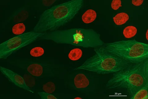
Before a cell divides, a dynamic structure helps sort its chromosomes. A new set of frozen-cell images examines how many short filaments can work together inside that structure.
- Cryo-electron tomography reconstructs local three-dimensional structure from frozen samples.
- The paper reports an approximately 8-nanometre wall-to-wall spacing in its structural analysis.
- The number of reconstructed image series is not the number of independent spindles.
- Paper date
- 29 October 2026
- Material
- Human cell spindles in frozen samples
- Focus
- Filament spacing, orientation and motor activity
Original publication: 29 Oct 2026 · The date above refers to this brief.
The spindle count is smaller than the image-series count
spindles in the Fig. 2 structural comparison
Read the cell-division experiment at three levels
Prepare
Freeze and image a defined laboratory sample.
Reconstruct
Map local filaments and their orientations.
Compare
Relate local organisation to spindle shape and motor conditions.
Why the spindle matters
During cell division, the mitotic spindle helps align and separate chromosomes. It contains microtubules, filament-like structures with directional ends, as well as molecular motors that can interact with them. A spindle looks organised at a large scale, but individual filaments can be much shorter than the structure itself.
Source 1 ↗What the frozen view adds
Conway and colleagues used cryo-electron tomography to reconstruct local microtubule arrangements in human cell spindles. The method provides three-dimensional structural information from frozen material. A reconstruction is a snapshot of the prepared sample; by itself it is not a movie showing every filament moving during division.
Source 1 ↗The eight-nanometre observation
The abstract reports a characteristic spacing of about 8 nanometres from wall to wall between neighbouring microtubules. The paper also examines local density and the orientation of filament ends. An observed spacing supports a structural description under the tested conditions; it does not mean every pair in every cell has exactly the same separation.
Source 1 ↗Why one pole is a useful comparison
The researchers also examined monopolar structures with one pole while motor activity remained present. Comparing local filament organisation in these samples and conventional metaphase spindles helps separate a local arrangement from the overall two-pole shape. Additional perturbations address the roles of the Eg5 and dynein motors.
Source 1 ↗Do not confuse images with spindles
The Fig. 2 comparison reports 24 tilt series from 11 metaphase spindles and 51 tilt series from 18 monopolar spindles. Several reconstructed series can come from the same spindle. These are laboratory structures and image series, not 24 and 51 independent people.
Source 1 ↗What a follow-up could add
Independent studies could test how well the observed local organisation generalises across cell types and how structures change over time. Live imaging and frozen structural snapshots answer complementary questions and have different limits. Our nuclear-pore guide explores a similar distinction between a structural map and a movie of movement.
Separate structural units before comparing numbers
Selected accepted primary PDF: abstract and introduction on pp. 1–2, structural comparison and Fig. 2 caption on pp. 4–5. Fig. 2 page was visually inspected. The accepted article may be edited. Publisher HTML metadata and abstract also checked. Supplements, source-data files, code and clinical records were not independently audited. No publisher figure reproduced.
01What does eight nanometres measure?
- What was observed
- The abstract reports characteristic wall-to-wall spacing between microtubules.
- Where the conclusion stops
- It is not the diameter of the spindle or a patient measurement.
Source 1 · Accepted PDF p. 1; abstract
02How many structures contributed to Fig. 2?
- What was observed
- The figure caption identifies 11 metaphase and 18 monopolar spindles.
- Where the conclusion stops
- The associated 24 and 51 tilt series are nested image series, not people.
Source 1 · Accepted PDF p. 5; Fig. 2 caption
Numbers you can inspect
| Measure | Value & unit | Origin & method |
|---|---|---|
| Characteristic wall-to-wall spacing | 8 nanometres, approximately | Reported Reported local microtubule spacing, not the whole spindle. Source 1 · PDF p. 1; abstract |
| Metaphase tilt series | 24 tilt series | Reported Image series contributed by 11 metaphase spindles. Source 1 · PDF p. 5; Fig. 2 caption |
| Metaphase spindles | 11 spindles | Reported Laboratory structures, not people. Source 1 · PDF p. 5; Fig. 2 caption |
| Monopolar tilt series | 51 tilt series | Reported Image series contributed by 18 monopolar spindles. Source 1 · PDF p. 5; Fig. 2 caption |
| Monopolar spindles | 18 spindles | Reported Laboratory structures, not people. Source 1 · PDF p. 5; Fig. 2 caption |
Compare the actual experiments
These studies answer different questions. Read the unit and endpoint before comparing results.
| Study | Unit & setting | Readout | Interpretation boundary |
|---|---|---|---|
| Metaphase reconstruction Source 1 · PDF p. 5; Fig. 2 caption | 24 tilt series from 11 human-cell spindles | Local microtubule organisation | A tilt series is nested within a spindle. |
| Monopolar comparison Source 1 · PDF p. 5; Fig. 2 caption | 51 tilt series from 18 one-pole structures | Local spacing and orientation | Overall shape and local organisation are different measures. |
The export includes claims, available numbers, methods and source locations. It contains our reading notes and published summaries; it is not raw participant data or an independent reanalysis.
Evidence update · 31 Oct 2026
First publication. Reported observations, arithmetic and proposed follow-up questions are separated. No raw-data reanalysis. AI source check; no human editorial or clinical review.
Three scales of a spindle study
| Reading question | Evidence to retain | What remains open |
|---|---|---|
| Local structure | Spacing and filament orientation in a reconstruction | The history of every filament |
| Whole spindle | A two-pole or monopolar overall arrangement | The direction of every local interaction |
| Motor experiment | Changes when a specified motor condition is altered | A treatment effect in people |
| Sample count | Tilt series nested within imaged spindles | One independent person per image |
Original Press-News reading framework. Follow-up questions are our interpretation, not experiments performed by the authors.
Your questions, answered
Is cryo-electron tomography a video?
No. It reconstructs structure from images of a frozen sample; time-course experiments need a different design.
Is 8 nanometres the whole spindle width?
No. The abstract describes an approximate wall-to-wall spacing between neighbouring microtubules.
Were 24 patients in the metaphase group?
No. The comparison reports 24 tilt series from 11 metaphase spindles.
Does the study establish a new cancer treatment?
No. It is a cell-structure study; a therapeutic claim would require separate evidence.
Limits of this interpretation
- A frozen reconstruction captures local structure and does not directly record the full history of spindle dynamics.
- Tilt series and spindles are different units; the comparison does not count patient outcomes.
- Findings in tested cell systems do not establish a cancer treatment or a personal risk.
- We did not independently segment the microtubules or reanalyse the raw reconstructions.
Sources & transparency
- Conway, Bobe, Zimyanin et al. (2026): CryoET imaging reveals that mitotic spindles are locally organized by microtubule density and Eg5 activity
Selected accepted primary PDF: abstract and introduction on pp. 1–2, structural comparison and Fig. 2 caption on pp. 4–5. Fig. 2 page was visually inspected. The accepted article may be edited. Publisher HTML metadata and abstract also checked. Supplements, source-data files, code and clinical records were not independently audited. No publisher figure reproduced. · Accessed 31 Oct 2026
DOI: 10.1038/s41467-026-78938-3
Prepared and source-checked with AI. Press-news Team is the collective publication byline, not a medical reviewer. No human editorial or clinical review has taken place. This educational article examines research methods and laboratory or observational findings, not individual diagnosis or treatment. We did not conduct these experiments or reanalyse raw data. Reported results, our interpretation and proposed future tests are separated. Source-access limits are recorded below. Photographs are illustrative.
Source check: AI source check — selected primary PDF results, captions and study boundaries
Clinical review: Not applicable to this educational guide
Suggest a correction