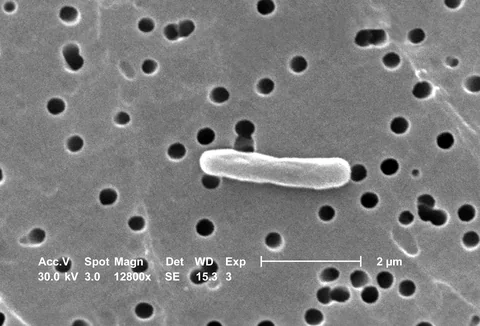
The surface of a bacterium is not uniform, and an image of its side need not reveal its end. A microscopy study examines how surface structure changes with species and orientation.
- The researchers used atomic force microscopy and optical measurements to examine bacterial surfaces in several laboratory strains.
- One comparison used 15 image areas from three biological repeats; a separate time series in Figure 4 shows one cell.
- The study describes research imaging and does not establish a new antibiotic or a clinical test.
- Paper date
- 9 November 2026
- Method
- Atomic force microscopy
- Selected image comparison
- 15 areas, 3 biological repeats
Original publication: 9 Nov 2026 · The date above refers to this brief.
From surface image to cautious conclusion
Observe
Measure a stable cell surface and use optical readouts alongside it.
Compare
Distinguish species, orientation, growth phase and sampled units.
Interpret
Keep unresolved features, selection effects and clinical relevance separate.
A surface is more than an outline
Atomic force microscopy uses a tiny probe to measure features of a surface. For living-cell imaging, the sample must remain sufficiently stable to measure while still retaining relevant biological properties. The authors combine this technique with optical measurements to study several bacterial species under laboratory conditions.
Source 1 ↗A side and an end are different views
Rod-shaped cells positioned horizontally and vertically expose different parts of their surface. In the studied E. coli cells, the authors report differences in the appearance of cell ends between stationary and growing phases. Some features were unresolved in growing cells; that does not prove they were absent.
Source 1 ↗A viability proxy has limits
The researchers used cell-envelope integrity as a proxy for viability and also show an example of growth and division during imaging. In one E. coli comparison, they report viability typically at least 90% over two hours for methods that successfully retained cells. This statement is specific to the tested conditions and proxy; it is not a guarantee for every species, cell or later time point.
Source 1 ↗Fifteen areas, three repeats, one cell
For Figure 4’s selected attachment and viability measurements, the caption reports 15 image areas across three biological repeats. Another image time series in the same figure follows one cell. These observations have different units: a close-up of one cell can illustrate a process but cannot establish how often it occurs across a whole population.
Source 1 ↗What remains uncertain
Different surface-attachment approaches may select different cells from a population. The authors also note that some surface features have uncertain origins. A nanoscale image needs careful interpretation, especially when the contrast may arise indirectly from surrounding material rather than a direct view of a particular molecule.
Source 1 ↗Connection: more detail is not the same as more certainty
Our article on two-photon imaging distinguishes spatial resolution from localization precision. The instruments and samples are different, but both studies show why labels, measurement conditions and sampling must accompany an impressive image. Neither paper evaluates a patient-facing diagnostic test.
An image area and a biological repeat are not the same unit
Primary PDF: abstract, Results, Fig. 2 and Fig. 4 caption p. 7, Discussion. The Fig. 4 caption page was visually inspected. Publisher metadata and abstract also checked. Supplements, source-data files, code and clinical records were not independently audited. No publisher figure reproduced.
01How many independent repeats support the selected Figure 4 measurements?
- What was observed
- The caption reports three biological repeats with five image areas each, giving 15 areas.
- Where the conclusion stops
- The 15 areas are not 15 independent biological repeats.
Source 1 · Primary PDF Fig. 4 caption p. 7
02Does a missing pattern prove a structure is absent?
- What was observed
- The authors report that some patterns on growing-cell poles were unresolved.
- Where the conclusion stops
- Unresolved means the image did not distinguish the feature, not that it is proven absent.
Source 1 · Primary PDF Results and Fig. 5
Numbers you can inspect
| Measure | Value & unit | Origin & method |
|---|---|---|
| Image areas in Figure 4 comparison | 15 areas | Reported Five acquisition areas from each of three biological repeats. Source 1 · Primary PDF Fig. 4 caption p. 7 |
| Biological repeats in Figure 4 comparison | 3 repeats | Reported Independent biological repeats for the selected measurements. Source 1 · Primary PDF Fig. 4 caption p. 7 |
| Areas per biological repeat | 5 areas per repeat | Reported Caption reports five acquisition areas for each repeat. Source 1 · Primary PDF Fig. 4 caption p. 7 |
| Cells in a selected Figure 4 time series | 1 cell | Reported One illustrated cell; separate from the 15-area comparison. Source 1 · Primary PDF Fig. 4 caption p. 7 |
Compare the actual experiments
These studies answer different questions. Read the unit and endpoint before comparing results.
| Study | Unit & setting | Readout | Interpretation boundary |
|---|---|---|---|
| Attachment and viability comparison Source 1 · Primary PDF Fig. 4 caption p. 7 | 15 image areas across three biological repeats | Attachment and envelope-integrity proxy | Image areas are nested within biological repeats. |
| One illustrated time series Source 1 · Primary PDF Fig. 4 caption p. 7 | One bacterial cell | Surface features over time | An individual example cannot estimate population prevalence. |
The export includes claims, available numbers, methods and source locations. It contains our reading notes and published summaries; it is not raw participant data or an independent reanalysis.
Evidence update · 16 Nov 2026
First publication. Reported observations, arithmetic and proposed follow-up questions are separated. No raw-data reanalysis. AI source check; no human editorial or clinical review.
Four questions behind a bacterial image
| Reading question | What the paper reports | What remains open |
|---|---|---|
| Orientation | Sides and ends can expose different features | A single view does not show the complete cell |
| Viability | Cell-envelope integrity was used as a proxy | Not a guarantee of normal behaviour in all conditions |
| Sampling | 15 image areas across three biological repeats in one comparison | Areas are not 15 independent biological experiments |
| Time series | One cell is shown in a selected Figure 4 series | One example cannot determine population frequency |
Original Press-News reading framework. Follow-up questions are our interpretation, not experiments performed by the authors.
Your questions, answered
Is an unresolved feature necessarily absent?
No. The authors distinguish features that could not be resolved from demonstrated absence.
Were 15 independent biological repeats performed for the Figure 4 comparison?
No. Fifteen image areas came from three biological repeats.
Does the viability proxy guarantee normal behaviour of every cell?
No. Cell-envelope integrity is one proxy under specific conditions.
Did the study demonstrate an antibiotic treatment?
No. It reports methods and observations of bacterial surfaces in laboratory models.
Limits of this interpretation
- The work studies selected laboratory strains and imaging conditions.
- Cell-envelope integrity is a proxy for viability, not a complete measure of cell health.
- Attachment can select a subset of the population; individual images are not prevalence estimates.
- An unresolved feature is not necessarily absent; some structures have uncertain origins.
Sources & transparency
- Scheeder et al. (2026): Nanometer-resolution imaging of living bacteria across species and cellular orientations
Primary PDF: abstract, Results, Fig. 2 and Fig. 4 caption p. 7, Discussion. The Fig. 4 caption page was visually inspected. Publisher metadata and abstract also checked. Supplements, source-data files, code and clinical records were not independently audited. No publisher figure reproduced. · Accessed 16 Nov 2026
DOI: 10.1038/s41467-026-78975-y
Prepared and source-checked with AI. Press-news Team is the collective publication byline, not a medical reviewer. No human editorial or clinical review has taken place. This educational article examines research methods and laboratory or observational findings, not individual diagnosis or treatment. We did not conduct these experiments or reanalyse raw data. Reported results, our interpretation and proposed future tests are separated. Source-access limits are recorded below. Photographs are illustrative.
Source check: AI source check — selected primary PDF results, captions and study boundaries
Clinical review: Not applicable to this educational guide
Suggest a correction