Every cell nucleus exchanges material with its surroundings through tiny gateways. A new study looks at how flexible proteins inside those gates move in living cells and in simplified laboratory systems.
- The authors used fluorescence measurements to study apparent movement in a nuclear-pore protein in living cells.
- A sugar modification and the transport protein importin-β influenced the behaviour of the studied system.
- Measurements in cells, purified protein mixtures and simulations answer related but different questions.
- Paper date
- 11 November 2026
- Focus
- Nuclear pore proteins
- Models
- Cells, purified proteins and simulations
Original publication: 11 Nov 2026 · The date above refers to this brief.
One figure, three groups of cells
cells in the Figure 4C motion analysis
Compare the settings
Measure
Estimate local motion in living cells.
Reconstruct
Study selected proteins in a simplified mixture.
Model
Test interpretations against imaging and simulations.
What is a nuclear pore?
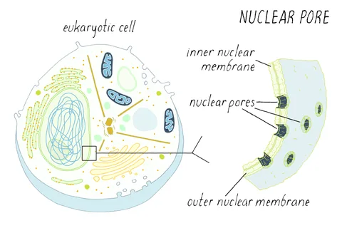
The nuclear envelope separates the nucleus from the rest of a cell. Nuclear pores are gateways through this envelope; their protein-rich interior helps control molecular traffic. The study focuses on a flexible region of a pore protein called NUP98 and asks how its motion depends on the surrounding cellular environment.
Source 1 ↗Cells and simplified mixtures behaved differently
The authors report liquid-like apparent segmental motion of the studied protein region in living cells. In reconstructed condensates of purified protein, the behaviour changed over time towards a more solid-like state. This comparison illustrates why a simplified mixture may not reproduce everything present in a living pore.
Source 1 ↗Two influences in the experiment
The researchers examined the effects of a sugar modification, O-GlcNAc, and a transport protein called importin-β. In their systems both influenced protein dynamics. Reducing the modification in cells was associated with slower apparent motion and transport; additional importin-β partly restored the measured responses under the tested conditions.
Source 1 ↗Why one figure has several sample counts
For one motion measurement in Figure 4C, the caption reports 80 control cells, 109 cells in the reduced-modification condition and 74 in the combined condition, across three independent experiments. A separate transport assay in the same figure has different cell counts. These numbers are cells in selected experiments, not patients, pores or independent clinical trials.
Source 1 ↗What was observed and what was modelled?
The study combines live-cell fluorescence measurements, experiments on purified protein assemblies, fixed-cell imaging and simulations. Simulations can test whether a proposed arrangement is consistent with assumptions; they are not a direct movie of every molecule in every living pore. The authors also use imaging as supporting evidence for spatial organisation.
Source 1 ↗Connecting a map with motion
Our earlier nuclear-pore map article explains why a distribution of molecular positions is not itself a film of movement. This new work adds measurements of apparent motion and transport, while still requiring readers to distinguish measured signals from the models used to interpret them.
Do not combine counts from different assays
Primary PDF: abstract and Results, Fig. 3 on p. 6 and Fig. 4 caption on p. 8. Selected figures and caption pages were visually inspected. Publisher metadata and abstract also checked. Supplements, source-data files, code and clinical records were not independently audited. No publisher figure reproduced.
01What do the three Figure 4C group sizes count?
- What was observed
- The caption reports 80, 109 and 74 cells across three independent experiments.
- Where the conclusion stops
- They count cells for one motion analysis, not people or clinical outcomes.
Source 1 · Primary PDF Fig. 4C caption p. 8
02Was every spatial detail directly filmed in living pores?
- What was observed
- The work combines live-cell signals, purified systems, fixed-cell imaging and simulations.
- Where the conclusion stops
- Simulated organisation and a fixed image are not a complete live molecular movie.
Source 1 · Primary PDF Results and Fig. 3 p. 6
Numbers you can inspect
| Measure | Value & unit | Origin & method |
|---|---|---|
| Control group | 80 cells | Reported Figure 4C motion analysis; across three experiments. Source 1 · Primary PDF Fig. 4C caption p. 8 |
| Reduced-modification group | 109 cells | Reported Figure 4C motion analysis; across three experiments. Source 1 · Primary PDF Fig. 4C caption p. 8 |
| Combined-condition group | 74 cells | Reported Figure 4C motion analysis; across three experiments. Source 1 · Primary PDF Fig. 4C caption p. 8 |
| Independent experiments | 3 experiments | Reported The three Figure 4C cell groups are reported across three experiments. Source 1 · Primary PDF Fig. 4C caption p. 8 |
Compare the actual experiments
These studies answer different questions. Read the unit and endpoint before comparing results.
| Study | Unit & setting | Readout | Interpretation boundary |
|---|---|---|---|
| Live-cell motion assay Source 1 · Primary PDF Fig. 4C caption p. 8 | Cells in Figure 4C | Fluorescence-derived apparent segmental motion | Cell counts and experimental replicates are different units. |
| Spatial analysis Source 1 · Primary PDF Fig. 3 p. 6 | Fixed-cell imaging and simulations | Predicted and observed localisation patterns | A fixed image or simulation is not a complete live trajectory. |
The export includes claims, available numbers, methods and source locations. It contains our reading notes and published summaries; it is not raw participant data or an independent reanalysis.
Evidence update · 13 Nov 2026
First publication. Reported observations, arithmetic and proposed follow-up questions are separated. No raw-data reanalysis. AI source check; no human editorial or clinical review.
Four ways to examine a nuclear pore
| Reading question | What the paper reports | What remains open |
|---|---|---|
| Live-cell fluorescence | Apparent local protein motion in cells | A measured signal still requires a model of its meaning |
| Purified protein mixture | Behaviour in a simplified assembly | It lacks parts of the cellular environment |
| Fixed-cell imaging | Spatial localisation of selected components | A fixed image is not a live movie |
| Simulation | Predictions under specified assumptions | Model agreement does not prove every detail |
Original Press-News reading framework. Follow-up questions are our interpretation, not experiments performed by the authors.
Your questions, answered
Did the researchers study a treatment?
No. This is basic research into how selected nuclear-pore proteins behave.
Does a liquid-like protein region mean the whole pore is liquid?
No. The paper describes apparent motion of selected flexible protein regions, not a claim that every part of the pore is a liquid.
What do 80, 109 and 74 count?
They are cells in three conditions for one Figure 4C motion analysis, across three independent experiments.
Are the simulations direct measurements?
No. They are computational models used alongside experiments to test interpretations.
Limits of this interpretation
- The work studies selected proteins and experimental cell systems, not a clinical intervention.
- Different figures use different sample sizes and assays.
- A fluorescence-derived timescale is an inferred measure of local motion, not a direct recording of each molecule’s complete path.
- Simulations and reconstructed condensates simplify the cellular environment; no raw-data reanalysis was performed.
Sources & transparency
- Yu, Heidari, Palacio-Rodriguez et al. (2026): Glycosylation and importins liquefy the FG barrier in live nuclear pores
Primary PDF: abstract and Results, Fig. 3 on p. 6 and Fig. 4 caption on p. 8. Selected figures and caption pages were visually inspected. Publisher metadata and abstract also checked. Supplements, source-data files, code and clinical records were not independently audited. No publisher figure reproduced. · Accessed 13 Nov 2026
DOI: 10.1038/s41586-026-11258-0
Prepared and source-checked with AI. Press-news Team is the collective publication byline, not a medical reviewer. No human editorial or clinical review has taken place. This educational article examines research methods and laboratory or observational findings, not individual diagnosis or treatment. We did not conduct these experiments or reanalyse raw data. Reported results, our interpretation and proposed future tests are separated. Source-access limits are recorded below. Photographs are illustrative.
Source check: AI source check — selected primary PDF results, captions and study boundaries
Clinical review: Not applicable to this educational guide
Suggest a correction