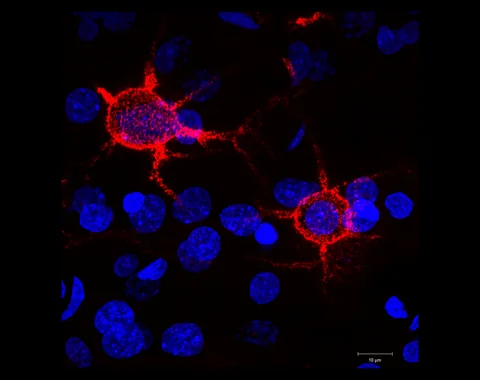
Pain has sensory and emotional dimensions that are difficult to separate experimentally. Researchers studied how selected brain cells respond after nerve injury in mice.
- The study combines brain-cell imaging and behavioural assays in mouse models, not a clinical trial.
- In one male-mouse analysis, the proportion of imaged neurons responding to more than one stimulus type rose from 2.8% to 21.2% after injury.
- That comparison concerns neurons from three male mice and is not a percentage of people whose pain changed.
- Paper date
- 16 November 2026
- Model
- Nerve injury in mice
- Selected measure
- Responses of imaged neurons
Original publication: 16 Nov 2026 · The date above refers to this brief.
A male-mouse neuronal response measure
percent of imaged neurons
From a mouse model to a cautious conclusion
Measure
Record selected neuronal responses before and after the experimental injury.
Compare
Consider sham controls, sex-specific analyses and behavioural readouts.
Interpret
Test generalization separately before drawing conclusions about people.
What were researchers trying to separate?
The authors examined a brain region in mice that participates in processing sensory and behavioural information. They combined measurements of activity in selected inhibitory neurons with behavioural experiments after nerve injury. These tools address particular features of the mouse model; they cannot directly measure another person’s subjective pain.
Source 1 ↗A change in a selected cell population
The paper reports that, in male mice, the proportion of imaged inhibitory neurons responding to more than one type of stimulus rose from 2.8% before injury to 21.2% afterward. Our subtraction gives an 18.4 percentage-point difference. This describes the reported neuronal measure, not an 18.4% change in a pain score.
Source 1 ↗How many animals were behind the images?
For the broader Figure 1 injury comparison, the caption reports 536 neurons from six mice; the sham comparison reports 726 neurons from seven mice. The injury-group sex breakdown includes 275 neurons from three male mice and 261 neurons from three female mice. The male-specific percentage should not be generalized to all animals or people.
Source 1 ↗Activity and behaviour are different readouts
The researchers also investigated how different neural cell classes relate to selected sensory and behaviour-based outcomes. These assays are useful for testing ideas about a circuit, but a behavioural proxy in a mouse does not capture every sensory or emotional aspect of human pain.
Source 1 ↗Why the control matters
A sham comparison helps researchers interpret changes associated with the injury procedure and repeated testing. The paper reports different patterns in injury and sham groups. Both the choice of model and the small numbers of animals in particular panels matter when deciding how broadly the result applies.
Source 1 ↗A question for future work
Independent replication, broader animal sampling and carefully designed human studies would be needed to learn how the proposed circuits relate to human conditions. This study does not identify an established therapy or support changing pain care. Our earlier microscopy article explains why details of the imaging method matter when interpreting a biological signal.
The unit is a neuron in a mouse, not a person
Primary PDF: abstract, Results and Fig. 1 caption p. 2, and selected subsequent Results. The Results and caption page was visually inspected. Publisher metadata and abstract also checked. Supplements, source-data files, code and clinical records were not independently audited. No publisher figure reproduced.
01Did 21.2% of patients improve?
- What was observed
- The paper reports 21.2% of imaged neurons in a male-mouse post-injury analysis responded to more than one modality.
- Where the conclusion stops
- The percentage is not a patient outcome or measure of treatment benefit.
Source 1 · Primary PDF Results p. 2
02Were 536 independent mice measured?
- What was observed
- The Figure 1 caption reports 536 neurons from six injured mice.
- Where the conclusion stops
- Neurons are nested within animals; the male subgroup contains three mice.
Source 1 · Primary PDF Fig. 1 caption p. 2
Numbers you can inspect
| Measure | Value & unit | Origin & method |
|---|---|---|
| Injury-group neurons | 536 neurons | Reported Imaged neurons from six mice in the selected Figure 1 comparison. Source 1 · Primary PDF Fig. 1 caption p. 2 |
| Injury-group mice | 6 mice | Reported Animal count for the Figure 1 injury group. Source 1 · Primary PDF Fig. 1 caption p. 2 |
| Sham-group neurons | 726 neurons | Reported Imaged neurons from seven sham mice. Source 1 · Primary PDF Fig. 1 caption p. 2 |
| Sham-group mice | 7 mice | Reported Animal count for the Figure 1 sham group. Source 1 · Primary PDF Fig. 1 caption p. 2 |
| Male-mouse pre-injury proportion | 2.8 percent of neurons | Reported Reported proportion responding to multiple stimulus modalities. Source 1 · Primary PDF Results p. 2 |
| Male-mouse post-injury proportion | 21.2 percent of neurons | Reported Reported proportion responding to multiple stimulus modalities. Source 1 · Primary PDF Results p. 2 |
| Reported proportion difference | 18.4 percentage points | Calculated 21.2 minus 2.8, using reported rounded percentages. Source 1 · Primary PDF Results p. 2 |
Compare the actual experiments
These studies answer different questions. Read the unit and endpoint before comparing results.
| Study | Unit & setting | Readout | Interpretation boundary |
|---|---|---|---|
| Neuronal imaging Source 1 · Primary PDF Fig. 1 caption p. 2 | Neurons nested within mice | Responses to stimulus modalities | Not a patient pain score. |
| Mouse behaviour experiments Source 1 · Primary PDF Results and Fig. 3 | Mouse groups in separate panels | Sensory and behaviour-based readouts | A proxy does not reproduce every aspect of human pain. |
The export includes claims, available numbers, methods and source locations. It contains our reading notes and published summaries; it is not raw participant data or an independent reanalysis.
Evidence update · 17 Nov 2026
First publication. Reported observations, arithmetic and proposed follow-up questions are separated. No raw-data reanalysis. AI source check; no human editorial or clinical review.
Keep the readouts and units apart
| Reading question | What the paper reports | What remains open |
|---|---|---|
| Imaging | 536 neurons from six injured mice in Figure 1 | Neurons are nested within animals |
| Sham comparison | 726 neurons from seven mice in the same figure | A separate control group |
| Male injury subgroup | 275 neurons from three mice | A subgroup, not all animals |
| Behavioural assays | Selected mouse responses in later experiments | Not a clinical pain rating |
Original Press-News reading framework. Follow-up questions are our interpretation, not experiments performed by the authors.
Your questions, answered
Does 21.2% refer to patients?
No. It is a reported proportion of imaged neurons in a male-mouse analysis.
Were 536 independent animals imaged?
No. The caption reports 536 neurons from six injured mice.
Were female mice included in the imaging comparison?
Yes. The Figure 1 injury group includes male and female subgroups, but the 2.8% to 21.2% result was reported for males.
Does the study establish a new pain treatment?
No. It reports experimental findings in mouse models, not a clinical treatment trial.
Limits of this interpretation
- The work uses experimental mouse models and selected neuronal populations.
- The 2.8% and 21.2% comparison is reported for male mice, with three male mice in the figure’s injury subgroup.
- Neuronal responses and behavioural assays are not direct measures of a person’s subjective experience.
- Individual figure panels have different sample sizes and outcomes.
Sources & transparency
- Gan, Oswald, Dou et al. (2026): Distinct prelimbic microcircuits control sensory and affective dimensions of neuropathic pain
Primary PDF: abstract, Results and Fig. 1 caption p. 2, and selected subsequent Results. The Results and caption page was visually inspected. Publisher metadata and abstract also checked. Supplements, source-data files, code and clinical records were not independently audited. No publisher figure reproduced. · Accessed 17 Nov 2026
DOI: 10.1038/s41593-026-02498-4
Prepared and source-checked with AI. Press-news Team is the collective publication byline, not a medical reviewer. No human editorial or clinical review has taken place. This educational article examines research methods and laboratory or observational findings, not individual diagnosis or treatment. We did not conduct these experiments or reanalyse raw data. Reported results, our interpretation and proposed future tests are separated. Source-access limits are recorded below. Photographs are illustrative.
Source check: AI source check — selected primary PDF results, captions and study boundaries
Clinical review: Not applicable to this educational guide
Suggest a correction