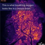
Imaging detects transient “hypoxic pockets” in the mouse brain
Using a bioluminescent oxygen indicator, Felix Beinlich and colleagues discovered a spontaneous, spatially defined occurrence of “hypoxic pockets” in the mouse brain. Their technique offers a way to learn more about brain oxygen tension (pO2), a measure of oxygen delivery and demand in brain tissue that changes dynamically but is not well understood. The findings could have implications for how rest and exercise affect pO2 in the human brain, including the role of these activities in conditions such as dementia, Beinlich et al. suggest. The researchers used a genetically encoded bioluminescent oxygen indicator called Green NanoLuc in mouse cortical astrocytes to ...