Novel calibration procedure for super-resolution brain imaging
A simple and robust procedure corrects a systematic error in microscopy, enabling precise imaging of biological tissue at greater depth
Light--and all waves--can bend around the corners of obstacles found along its path. Because of this phenomenon, called diffraction, it is impossible to focus light onto a spot that is smaller than half its wavelength. In other words, the highest resolution one can theoretically achieve using an optical microscope is approximately 250 nm, a barrier called the diffraction limit. Unfortunately, this resolution is not enough for observing fine cellular structures, such as those found in neurons.
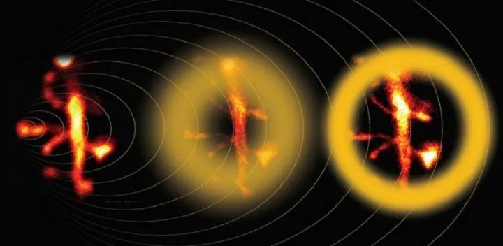
Over more than a century, microscopists were hamstrung by this classic barrier until the invention of super-resolution fluorescence microscopy. One particularly powerful approach was developed in the late 1990s and coined stimulated-emission depletion (STED) microscopy. This technique requires the target sample to contain fluorophores, which are compounds that absorb light at one wavelength and then re-emit it at a longer one. In the simplest version of STED microscopy, fluorophores are excited in a circular spot by irradiation with a diffraction-limited focused laser. Then, a donut-shaped portion around the spot is irradiated with less-energetic light--the depletion beam--which switches off the fluorescence by the process of stimulated emission. Thus, the net effect is that only the fluorophores in the center of the donut re-emit photons, and because that area can be made arbitrarily small, this allows for super-resolution microscopy.
Although STED microscopy was a true breakthrough for observing the morphology of live neurons at higher resolution, there is still room for improvement. END