Spallation Neutron Source accelerator achieves world-record 1.7-megawatt power level to enable more scientific discoveries
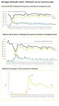
The Spallation Neutron Source at the Department of Energy's Oak Ridge National Laboratory set a world record when its particle accelerator beam operating power reached 1.7 megawatts, substantially improving on the facility’s original design capability.
The accelerator’s higher power provides more neutrons for researchers who use the facility to study and improve a wide range of materials for more efficient solar panels, longer–lasting batteries and stronger, lighter materials for transportation. The achievement marks a new operational milestone for ...